TIBIAL INTERCONDYLAR EMINENCE
FRACTURE
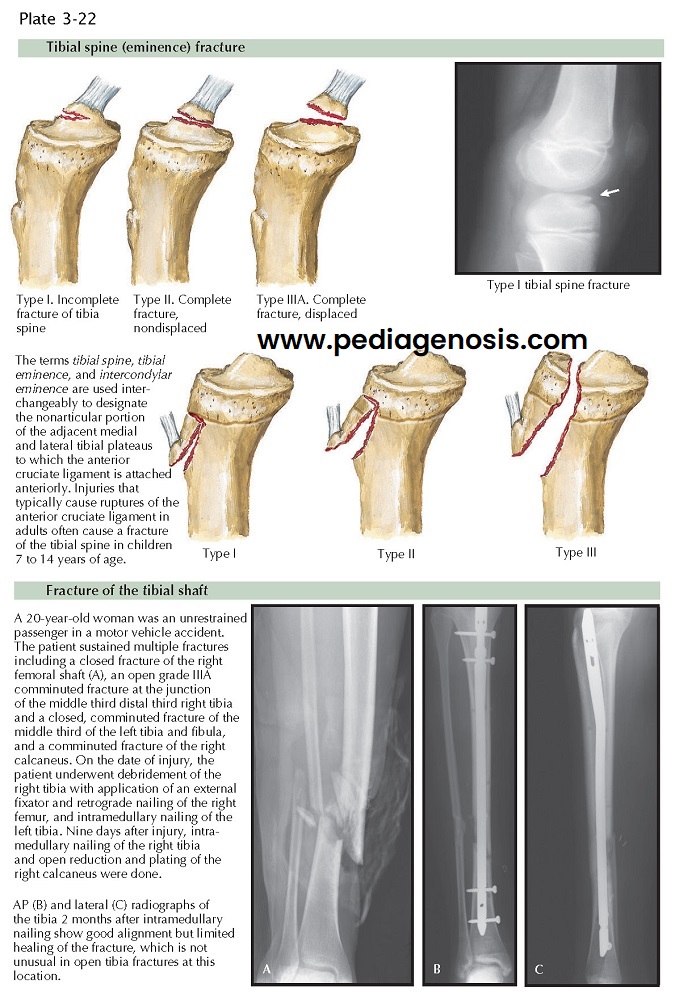
Fracture of the intercondylar eminence (tibial spine) indicates partial or complete detachment of the ACL from the tibia and is most commonly found in children. This fracture is usually caused by hyperextension of the knee or a sudden twisting motion. Forceful traction resulting from a direct blow to the distal femur on a flexed knee may also result in this fracture. If the fracture is displaced, the loose fragment may block motion and cause severe swelling and hemarthrosis. Type I fracture of the tibial spine is an incomplete fracture, whereas type II is complete but nondisplaced. Type III fractures are described as type IIIA (complete and displaced) and type IIIB (complete, displaced, and rotated out of position).
Nondisplaced fractures and those that reduce
anatomically when the knee is in full extension may be treated by casting or
rigidly bracing the knee in extension or in 20 degrees of flexion to increase
relaxation of the ACL. Union usually occurs in 5 to 6 weeks, after which the
patient begins active range-of-motion exercises and rehabilitation of the
quadriceps femoris and hamstring muscles.
Surgery is indicated for displaced type IIIA and type IIIB fractures of the tibial spine that cannot be adequately reduced with closed methods. Because this is an intra-articular fracture, anatomic reduction is required for return of knee function. Any mechanical block to full extension is also an indication for surgery. Reduction of the fracture may be blocked if the anterior horn of either meniscus is interposed between the displaced tibial spine and its bed. During surgery, all soft tissue is removed from the fracture site and the tibial spine is replaced in its anatomic position and fixated with sutures or a screw. This can be accomplished arthroscopically or through an arthrotomy. Because this fracture is most commonly found in the skeletally immature patient, appropriate attention must be paid to open physes during any surgical procedure. Adequate and rigid internal fixation allows the patient to regain motion quickly while the injured knee is protected in a brace. Once adequate fracture healing has occurred, postoperative rehabilitation protocols are similar to those undertaken in patients who have undergone ACL reconstruction.


