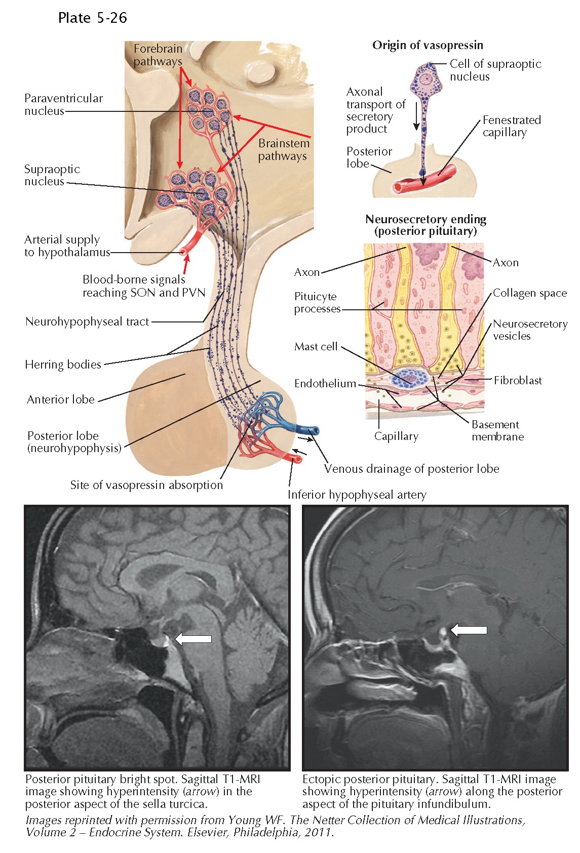
Posterior Pituitary Gland
The neurohypophysis, including the neural stalk and the posterior pituitary lobe, is an extension of the hypothalamus. It is embryologically derived from neural ectoderm. During development, magnocellular neurons of the supraoptic and paraventricular hypothalamic nuclei send their axons inferiorly to form the neurohypophysis. These axons terminate in the posterior lobe of the pituitary. In addition, a smaller number of parvocellular neurons from the same nuclei send off shorter axons, which end in the median eminence or infundibular stem. Pituicytes, which are glial cells, support these axon terminals. Two nonapeptide hormones are secreted from distinct axon terminals in the neurohypophysis, including antidiuretic hormone (vasopressin) and oxytocin. After secretion, these hormones enter neurohypophyseal capillaries and are carried via the inferior hypophyseal veins into the systemic circulation.
Oxytocin and
antidiuretic hormone (vasopressin) are made by distinct populations of large
(magnocellular) neurons in both the paraventricular (PVN) and supraoptic (SON)
nuclei, which release the hormones from their axons in the neurohypophysis. The
PVN also contains populations of smaller (parvicellular) neuroendocrine neurons
that produce corticotropin-releasing hormone, thyrotropin-releasing hormone, or
somatostatin, which are released into the hypothalamohypophyseal portal
circulation. Some parvicellular neurons in the PVN also make either oxytocin or
antidiuretic hormone, which are used as central neurotransmitters to control
the autonomic nervous system. Oxytocin and antidiuretic hormone are coded by
distinct genes, which code for precursor proteins that undergo
posttranslational cleavage into the hormone, a neurophysin protein, and a
carboxyterminal peptide called a copeptin. These are transported within
secretory granules (vesicles), traveling along neuronal axons until they reach
their respective axon terminals, where they are stored until secreted.
The presence of
secretory granules within the posterior pituitary lobe gives rise to a bright
signal on sagittal views of the pituitary on unenhanced, T1-weighted magnetic
resonance images, termed the “posterior bright spot.” This is present in 80% to
90% of healthy individuals. An ectopic posterior pituitary may be present at
the base of the hypothalamus or the pituitary stalk in some individuals, who
usually have intact posterior pituitary function. However, anterior pituitary
hypoplasia and variable anterior pituitary hormone deficiencies are often
present in these patients.
Release of
antidiuretic hormone or oxytocin occurs via exocytosis in response to action
potentials propagating along the respective axons. The release of antidiuretic
hormone and oxytocin is regulated by specific neural inputs to the hypothalamic
nuclei synthesizing these hormones, with glutamate representing a major
stimulatory neurotransmitter and γ-aminobutyric acid being
inhibitory. Both osmotic (increase in plasma osmolality, sensed by hypothalamic
osmoreceptors) and nonosmotic (significant decrease in effective arterial blood
volume, pain, nausea, certain medications) stimuli mediate antidiuretic hormone
release. Once secreted, antidiuretic hormone leads to increased water
permeability in the collecting ducts of the kidneys, resulting in water
reabsorption and urine concentration. Other antidiuretic hormone effects
include vasoconstriction, stimulation of glycogenolysis, and augmentation of
corticotropin release from the anterior pituitary.
Failure of
antidiuretic hormone synthesis and secretion is the cause of central diabetes
insipidus, which is characterized by passage of large volumes of dilute
urine, driving increased thirst. Dehydration and hypernatremia may generally
occur if access to water is limited in untreated patients. Although diverse
space-occupying lesions in the hypothalamus or within the sella may lead to
central diabetes insipidus, it may also be noted that pituitary adenomas only
rarely cause this condition preoperatively. The “posterior bright spot” is
generally absent on magnetic resonance imaging in patients with central
diabetes insipidus.
The release of oxytocin occurs in response to neural inputs during parturition, suckling, and intercourse. Animal data have suggested a role for oxytocin in propagation of labor and milk let-down during suckling. However, women who are deficient in oxytocin may experience normal labor and successful lactation, suggesting that the physiologic role of oxytocin in humans remains incompletely understood. A possible role of oxytocin in behavior is under study. There is also evidence in animals that oxytocin as a central neurotransmitter ay promote maternal behavior and social interaction.


