INJURY TO PELVIS
FRACTURE
WITHOUT DISRUPTION OF PELVIC RING
Injuries to the pelvis range from the most trivial to
the most life threatening. In general, the severity of an injury is determined
by the degree of disruption to the pelvic ring. Fortunately, many fractures
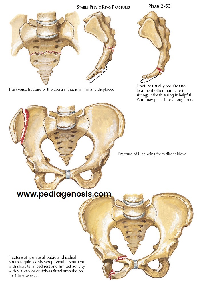
about the pelvis do not affect its integrity at all (see Plate 2-63).
 |
| STABLE PELVIC RING FRACTURES |
Avulsion
In athletes, avulsions of bone from the pelvis due to strong muscle contractions are relatively common. These fractures occur at the anterior superior iliac spine as a result of the strong pull of the sartorius muscle, at the anterior inferior iliac spine from the pull of the rectus femoris muscle, and at the ischial tuberosity from the forceful contraction of the hamstring muscles. Most fractures can be treated with activity modification and medications until pain ceases. Patients may gradually resume athletic activities, usually attaining complete functional recovery within 3 months.
Fracture of Iliac Wing
An isolated fracture of the iliac wing, or Duverney
fracture, is not uncommon, usually resulting from a direct compressive force to
the lateral aspect of the pelvis. The strong muscle attachments on the fragment
usually minimize its displacement; and although blood loss may be substantial,
shock is not common. Radiographic examination must determine whether the
acetabulum is involved or if the sacroiliac joint is disrupted. Management of
the fracture involves bed rest on a firm mattress until the patient is
comfortable enough to be mobilized, followed by gradual, protected weight
bearing until all symptoms resolve. Occasionally, an ileus develops that is not
due to abdominal visceral injury. This complication is usually relieved with
nasogastric suction and intravenous administration of fluids.
Fracture of Pubic or Ischial Ramus
An isolated fracture of a single pubic or ischial
ramus is a fairly uncommon injury; it usually occurs in elderly persons as a
result of a fall. In this type of fracture, the pelvis remains extremely stable
because the obturator foramen is a rigid, bony circle. A fracture of a pubic or
ischial ramus must be differentiated from an impacted fracture of the femoral
neck because treatments differ markedly. A fracture of one pubic or ischial
ramus without visceral or vascular injury requires only bed rest until symptoms
abate sufficiently to allow progressive mobilization with a gradual increase in
weight bearing.
Fracture of Sacrum
Transverse fractures of the sacrum are usually caused
by direct impact and typically have a slightly anterior displacement. Diagnosis
is based on a history of injury and evidence of pain, swelling, and tenderness
over the posterior aspect of the sacrum. Neurologic dysfunction may occur,
evidenced by urinary retention or decrease in rectal tone. The examiner must
take extreme care during the rectal
examination, especially during palpation along the anterior surface of the
sacrum, to avoid converting a closed sacral fracture into an open fracture
through the rectum, which increases the risk of serious contamination of the
retroperitoneal space. If neurologic deficit is either absent or improving,
conservative treatment is indicated. If a neurologic lesion compromises bowel
or bladder function, surgical decompression
should be considered.
This fracture is usually caused by a direct blow to
the posterior aspect of the coccyx. Symptomatic management suffices, but
discomfort may persist for a long time.
FRACTURE OF ALL FOUR PUBIC RAMI
When the pelvic ring is injured at more than one site,
the potential for instability increases correspondingly. Fractures of all four
pubic rami disrupt the structural integrity of the anterior portion of the
pelvic ring (see Plate 2-64). This injury commonly results from a fall on the front of the pelvis
or from lateral compressive forces on the pelvic ring. In about one third of patients,
major trauma to the lower urinary tract occurs as well. Treatment focuses on
preventing any further displacement of the fracture fragments. Bed rest, with
the patient in a semi-sitting position to relax the abdominal musculature, is
followed by mobilization as soon as symptoms allow.
STRADDLE FRACTURE AND LATERAL COMPRESSION INJURY
LATERAL COMPRESSION INJURY
Double breaks in the pelvic ring are often due to
lateral compressive forces (see Plate 2-64). Most of these
fractures are stable because the forces cause impaction of the posterior pelvic
complex, leaving the posterior ligaments intact. If the force continues or
increases, the posterior sacroiliac ligaments may tear, producing an
instability in the injured hemipelvis.
This injury often includes fractures of the superior
and inferior pubic rami; therefore, the anterior and posterior injuries are on
the same side. When the patient is placed in the supine position, the displaced
hemipelvis often reduces spontaneously. Radiographs may reveal only minimal
displacement, but the examiner must remember that the degree of initial
deformation of the hemipelvis is not known, and significant visceral
injury may be present.
When a lateral compressive force is accompanied by a
rotational force, the anterior fracture often occurs on the side opposite the
posterior fracture; for example, an impacted fracture may occur on the left
side of the sacrum while fractures of the inferior and superior pubic rami
occur on the right side. The hemipelvis then displace’s superiorly and medially,
causing the leg to appear internally rotated and shortened. The position of the
hemipelvis generally remains stable, although the malrotation may compromise
future function. Correcting this rotational deformity may create instability when the impacted posterior injury is reduced.
ANTEROPOSTERIOR COMPRESSION FRACTURE
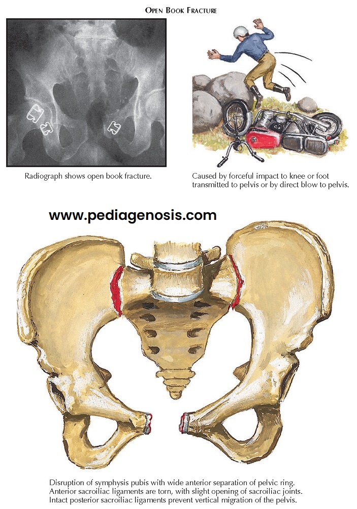
Anteroposterior compression, or open book, fractures
of the pelvis are usually caused by a forceful blow from in front of the pelvis
through the anterior superior iliac spines or by force applied to externally
rotated femurs (see Plate 2-65).
However, a blow from the rear against the posterior superior iliac spines may
produce a similar injury that is characterized by disruption of the pubic
symphysis and the anterior sacroiliac ligaments. Injuries range from an
isolated rupture of the pubic symphysis to complete separation of the pubic
symphysis accompanied by bilateral anterior subluxation of the sacroiliac joints.
Because the sacrospinal ligaments resist external rotation of the pelvis, a
separation of 2.5 cm or greater of the symphysis suggests that the sacrospinal
ligaments have ruptured or have avulsed bone from the sacrum or ischial spine.
Most important, the very strong posterior sacroiliac ligaments remain intact.
The open book fracture is therefore relatively stable, and treatment focuses on
closing the pelvic ring.
The pelvic ring can be closed with or without surgery.
In nonsurgical treatment, crossover slings are used to bring the two pelvic
halves together. In 3 to 4 weeks, when the soft tissues have healed
sufficiently, the pelvic slings are replaced with a mini spica cast and
ambulatory treatment can be initiated. If surgical treatment is chosen, ORIF is
performed, using a plate to stabilize the pubic symphysis. The planning of
treatment for this injury must be clearly distinguished from the treatment for
a vertical shear injury of the pelvis. Because the initial radiographs of both
injuries may appear similar, differentiation requires careful physical examination
and supplemental imaging, including CT of the posterior pelvic complex.
VERTICAL SHEAR FRACTURE
Vertical shear, or Malgaigne, fractures of the pelvis
result from severe trauma (see Plate 2-66). The force may cause unilateral or
bilateral injuries to the posterior pelvis and severe and sometimes
life-threatening injury to the soft tissue contents of the pelvic cavity. The
injury to the anterior pelvis may include disruption of the pubic symphysis
with or without fractures of two, three, or four pubic rami. The posterior
injuries may be fractures of the sacrum, dislocations of the sacroiliac joint,
fractures of the ilium, or fracture dislocations of the sacroiliac joint,
including fractures of the sacrum or the ilium.
Avulsion fractures, particularly of the ischial spine
or the transverse process of the fifth lumbar vertebra, also occur in major
disruptions of the posterior pelvis. Complete instability of the affected
hemipelvis indicates disruption of the sacrotuberal and sacrospinal ligaments.
Because vertical shear fractures are caused by considerable force, many involve
vital pelvic and abdominal tissues of the gastrointestinal, genitourinary,
vascular, and nervous systems.
If the posterior hemipelvis on one side remains
intact, the disrupted half can be brought to the intact side and stabilized.
When both ilia are disrupted from the sacrum, stabilization is much more
problematic. The aim of treatment soon after injury is to reapproximate the injured hemipelvis to the uninjured side using
closed manipulation. After anesthesia or analgesia with significant muscle
relaxation is obtained, the patient is positioned injured side up, with an
assistant supporting the legs. The examiner pushes the displaced ilium downward
and forward. If the reduction is successful, skeletal traction is often used to
maintain it until significant healing of soft tissue or bone occurs.
Because many Malgaigne fractures are grossly unstable,
manipulative reduction is largely unsuccessful, and either external or internal
fixation of the hemipelvis is needed. Fixation with an external fixation device
provides relative immobilization of the hemipelvis; the stability is greater
than that provided by manipulative reduction and subsequent traction. However,
attempts to attain and maintain anatomic reduction with
external fixators are usually
unsuccessful.
ORIF of both
displaced components provides the best stabilization of vertical shear
fractures. Two plates may be used to secure the disruptions in the pubic
symphysis and two bolts inserted to stabilize a longitudinal fracture of the
sacrum.
VASCULAR AND VISCERAL TRAUMA
Vascular and visceral injuries are the chief causes of
deaths associated with pelvic fractures. The likelihood of associated injury is
directly related to the severity of the fracture.
Pelvic fractures normally cause bleeding from injured
vessels of the pelvic marrow or from torn pelvic or lumbar arteries and veins
and formation of a hematoma. The extent of the bleeding depends on the severity
of the vascular and bone injuries, and the risk of death is directly linked to
the severity of the hemorrhage. Patients with double breaks in the pelvic ring
require transfusions twice as often as patients with single breaks or with
fractures of the acetabulum. Persistent severe hemorrhage may warrant emergent
investigation with transfemoral arteriography to identify bleeding sites and
selective embolization using blood, gelatin, or other such substances.
Arteriography may be used to identify injuries to large arteries. Military antishock
trousers may be used to stabilize the fracture and to help diminish blood loss.
Frequently, pelvic fractures cause injuries of the
lower urinary tract. Clues to such injuries include blood at the urethral
meatus or hematuria found on urinalysis. In men, rectal examination may reveal
a high-riding or free-floating prostate, which usually indicates injury to the
lower urinary tract. Urethrography is performed to determine whether the
anterior portion of the urethra is injured. If the urethrogram appears normal,
a catheter should be passed gently through the urethra into the bladder. The
catheter must not be forced. If this procedure is accomplished, further
radiographic studies can determine whether an intraperitoneal or an extra-peritoneal bladder injury is present.
 |
| VERTICAL SHEAR FRACTURE |
Most complete transections of the urethra are treated
by diverting the urine stream through a suprapubic catheter placed into the
bladder and, later, with repair or reconstruction of the urethra.
Intraperitoneal ruptures of the bladder are repaired with suprapubic drainage
to decompress the bladder. Depending on the extent of the tear, extraperitoneal
ruptures may not require surgical repair; adequate drainage of the bladder is necessary to allow the laceration to heal.
The examiner must search diligently for injuries of the lower gastrointestinal tract, especially about the rectum and the anus. Blood found on rectal examination suggests that the lower gastrointestinal tract has been lacerated by bone fragments, with possible severe contamination of the extraperitoneal pelvic space. If the rectum or anus is lacerated, standard treatment includes performing a washout of the distal rectal lumen and creating a diverting colostomy to minimize pelvic contamination. In women, a careful vaginal examination is performed to detect lacerations of the vagina, which could lead to contamination and infection of the pelvis.


