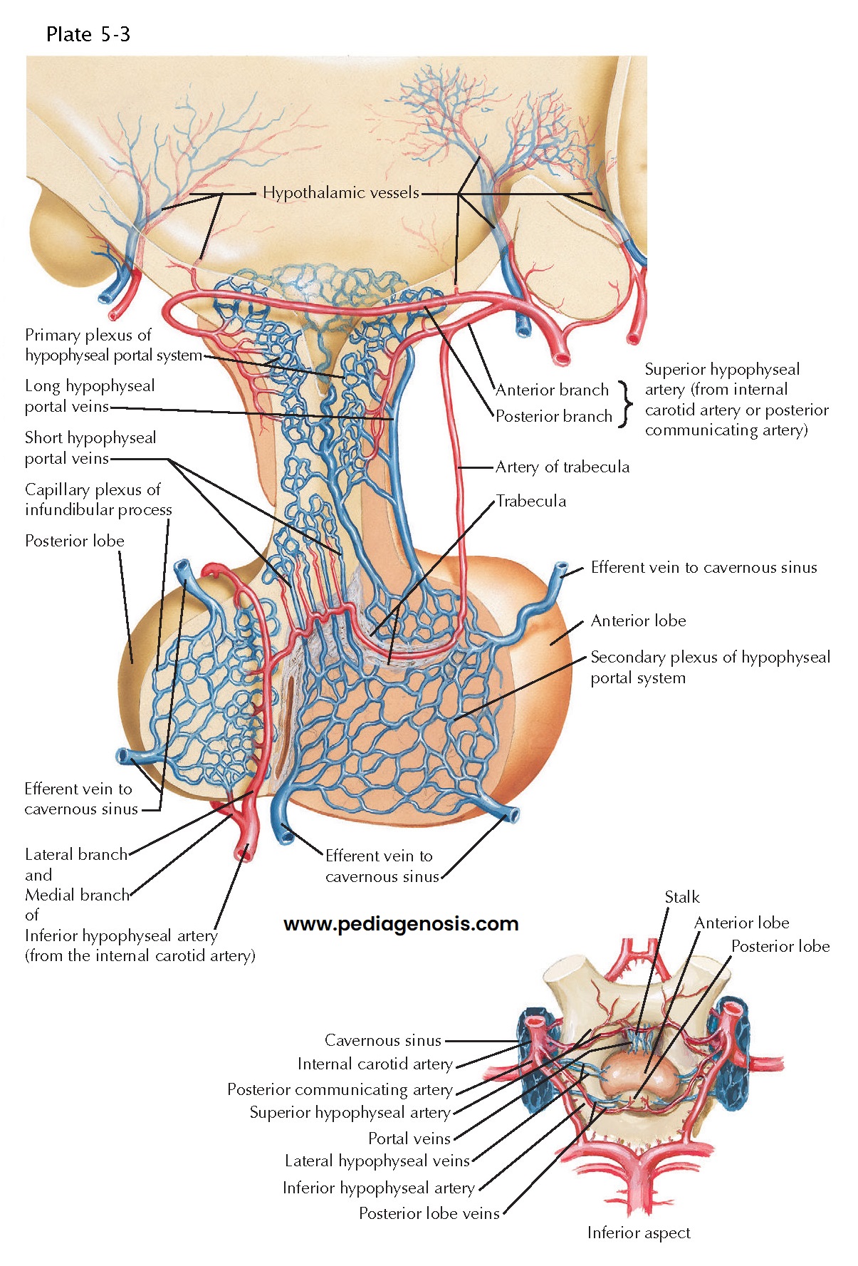
Blood Supply of the Hypothalamus and Pituitary Gland
The hypothalamus is what the circle of Willis encircles. The internal carotid artery runs through the cavernous sinus, which is just below the hypothalamus, and the site of its venous drainage. As the internal carotid artery emerges from the cavernous sinus, it ends in the middle cerebral artery laterally, the posterior communicating artery caudally, and the anterior cerebral artery rostrally. The anterior cerebral artery runs above the optic nerve, crosses the olfactory tract, and meets the anterior communicating artery in the midline before turning upward and back. The posterior communicating artery runs back to meet the posterior cerebral artery shortly after it emerges from the basilar artery. As a result, the hypothalamus is fed by small penetrating arteries that originate directly from the tributaries of the circle of Willis.
The anterior
part of the hypothalamus, above the optic chiasm, is supplied by arterial
feeding vessels from the anterior cerebral artery. These vessels densely
penetrate the basal forebrain just in front of the optic chiasm, giving it the
name the “anterior perforated substance.” The tuberal, or midlevel of the
hypothalamus, is fed mainly by small branches directly from the internal
carotid artery and the posterior communicating artery. Posteriorly, small
penetrating vessels from the posterior cerebral arteries running through the
inter-peduncular fossa give it the name “posterior perforated substance.” Many
of these small blood vessels supply the posterior part of the thalamus, but
some also provide blood to the posterior hypothalamus. The cell groups within
the hypothalamus are not uniformly supplied with blood vessels. The
paraventricular and supra-optic nuclei, which contain neurons that make the
vasoactive hormones oxytocin and vasopressin, have particularly rich capillary
networks.
The superior
hypophyseal artery is one of the branches derived from the internal carotid
artery. It supplies the pituitary stalk, where it breaks up into a series of
looplike capillaries in the median eminence and pituitary stalk. The
hypothalamic neurons that make pituitary releasing (and release-inhibiting)
hormones send axons that terminate on these loops, which, unlike most brain
capillaries, have fenestrations to permit easy penetration by these small
peptide hormones (see Plate 5-6). These capillaries drain into the hypophyseal
portal veins, which along with some branches of the inferior hypophyseal
artery, provide blood flow to the adenohypophysis or anterior pituitary gland.
The posterior pituitary gland is supplied almost entirely by the inferior
hypophyseal artery. Because most of the blood flow to the anterior pituitary
gland is from the portal system, it is possible, on occasions, for the gland to
outgrow its blood supply. This occurs mainly during pregnancy or can occur when
a pituitary adenoma, an otherwise benign tumor, becomes larger than can be
accommodated by the blood supply. At this point, there is infarction of the
pituitary, often with bleeding, which may become life threatening (pituitary
apoplexy). The typical presentation is sudden onset of dysfunction of cranial
nerve II, III, IV, or VI, with a severe headache that is generally localized
between the eyes, and often impaired consciousness.
Finally, the fenestrated capillary loops in the median eminence not only allow egress of hypothalamic-releasing hormones to the anterior pituitary gland, but also permit blood-borne substances to enter the brain. The hormone leptin, which is made by white adipose tissue during times of plenty, is believed to enter the brain via the median eminence to signal satiety to cell groups in the basal medial hypothalamus. There is another area of fenestrated capillaries along the anterior wall of the third ventricle, called the organum vasculosum of the lamina terminalis, which may allow entry of other hormones, such as angiotensin, which may be involved in thirst and water balance, and perhaps some cytokines that may play a role in the fever response. These regions are called circumventricular organs because they are around the edges of the ventricles. Another circumventricular organ, the area postrema, is found at the outflow of the fourth ventricle in the medulla and is probably involved in emetic reflexes based on blood-borne oxins or hormones, such as glucagon-like protein 1.


