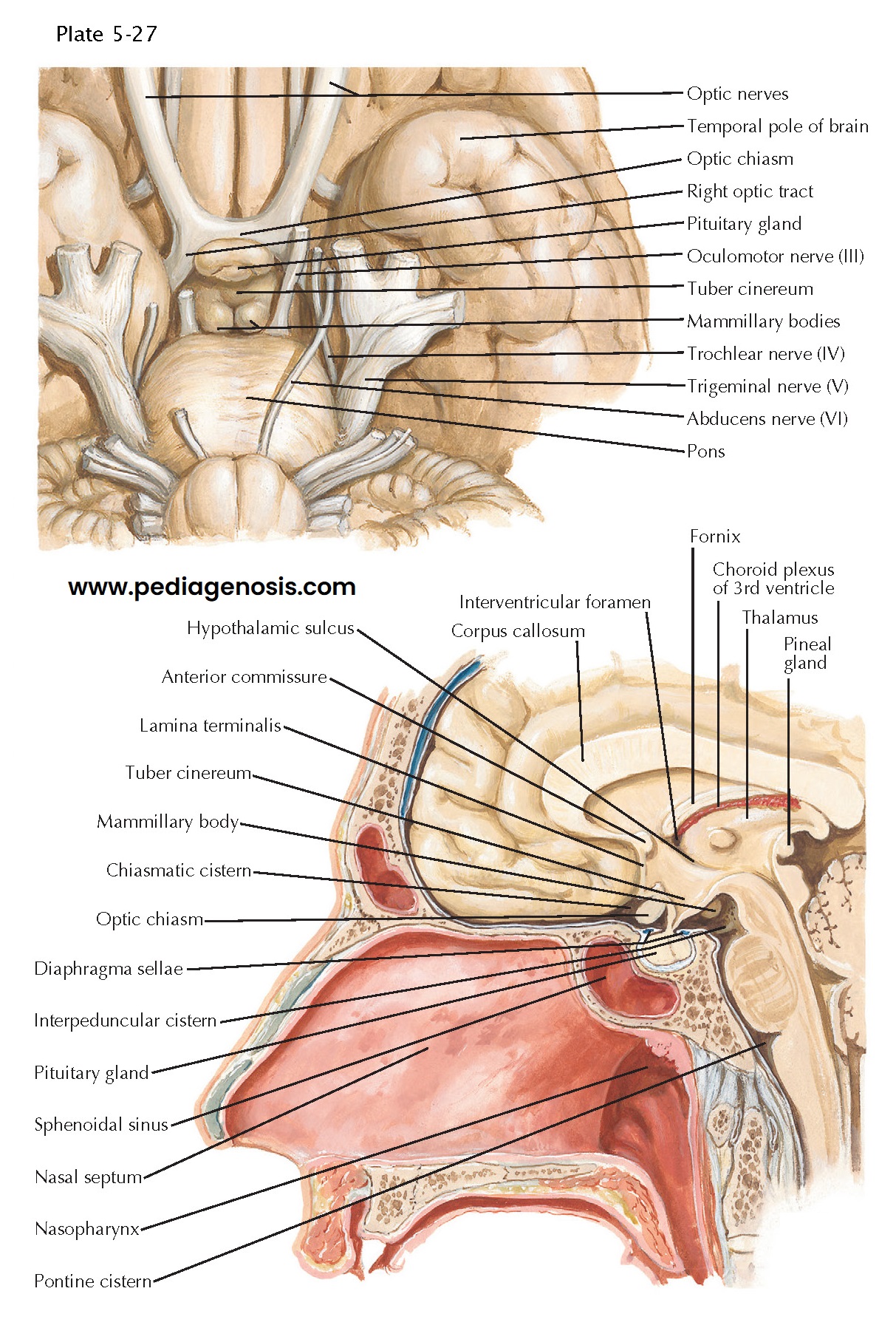
Anatomic Relationships of the Pituitary Gland
The pituitary gland resides in a depression (fossa) in the body of the sphenoid bone, termed the sella turcica. The tuberculum sellae forms the anterior wall of the sella, and the dorsum sellae forms its posterior wall. The pituitary is covered superiorly by a circular fold of dura mater, the diaphragma sellae. This sellar diaphragm is pierced by the pituitary stalk and the hypophyseal vessels. A fold of the arachnoid may herniate through the sellar diaphragm in some patients, thus extending the subarachnoid space within the sella (see MRI scan on the right in Plate 5-26 for an example). Chronic pulsatile pressure exerted by the cerebrospinal fluid may expand the sella, leading to the appearance of an enlarged, “empty sella,” which may be associated with hypopituitarism in some patients.
The optic
chiasm rests superiorly to the diaphragma sellae. Nerve fibers originating
in the nasal portion of each retina cross at the chiasm to the contralateral
side and join ipsilateral nerve fibers originating in the temporal portion of
each retina, which do not cross at the chiasm, to form each optic tract. The
anatomic relationship between the pituitary gland and the optic chiasm is
clinically important, because mass lesions within the pituitary may compress
either the chiasm or other portions of the optic apparatus, giving rise to a
variety of visual field defects. Specific places where the arachnoid separates
from the pia mater form cisterns filled with cerebrospinal fluid, including the
chiasmatic cistern and the interpeduncular cistern. These spaces
can be distorted by space-occupying sellar lesions growing superiorly from the
pituitary.
The hypothalamus
is located superiorly to the pituitary gland and is bounded between the optic
chiasm anteriorly, the caudal border of the mammillary bodies posteriorly,
and the hypothalamic sulcus superiorly. Distinct hypothalamic nuclei regulate
anterior pituitary function through the synthesis of several stimulating hormones
(growth hormone–releasing hormone, corticotropin-releasing hormone,
thyrotropin-releasing hormone, and gonadotropin-releasing hormone) and
inhibiting hormones (somatostatin and dopamine), released at neuronal axon
terminals present in the median eminence and the infundibulum.
These hormones are carried via the hypophyseal portal system to the
adenohypophysis, where they regulate hormone secretion in a specific manner.
The inter-relationships between the hypothalamus and the posterior pituitary
are detailed in Plate 5-26. Large lesions arising above the sella may impinge
on the hypothalamus, interfering with its functions.
The cavernous
sinuses are located laterally to the pituitary gland, and receive blood
from the pituitary via the hypophyseal veins. Each cavernous sinus contains
several important structures, including the cavernous portion of the
ipsilateral internal carotid artery, the oculomotor, trochlear, and
abducens nerves, as well as the first two divisions (ophthalmic and
maxillary) of the trigeminal nerve. Each of these nerves may be impinged
upon by space-occupying lesions arising in the sella that extend into the
cavernous sinus. Examples of such lesions include meningiomas,
chondrosarcomas and sellar metastases. Characteristically, pituitary
adenomas only rarely cause dysfunction of cranial nerves within the
cavernous sinuses, with the notable exception of adenomas undergoing
hemorrhagic necrosis (pituitary apoplexy). The circular sinus lies
between the pituitary gland and the underlying sphenoid bone in the sella,
forming interconnections between the two cavernous sinuses.
The thin sellar floor separates the pituitary gland from the underlying sphenoid sinus. The sellar floor can be expanded by slowly growing sellar masses, leading to remodeling of the sella, or eroded by sellar masses growing inferiorly. The close relationship between the sella, the sphenoid sinus, and the nasopharynx provides an important access route to pituitary surgeons. Using a trans-sphenoidal approach, many sellar masses can be resected with low morbidity.


