Anatomic
Relationships of the Hypothalamus
The hypothalamus is a small
area, weighing about 4 g of the total 1,400 g of adult brain weight, but it is
the only 4 g of brain without which life itself is impossible. The hypothalamus
is so critical for life because it contains the integrative circuitry that
coordinates autonomic, endocrine, and behavioral responses that are necessary
for basic life functions, such as thermoregulation, control of electrolyte and
fluid balance, feeding and metabolism, responses to stress, and reproduction.
 |
| ANATOMY AND RELATIONS OF THE HYPOTHALAMUS AND PITUITARY GLAND |
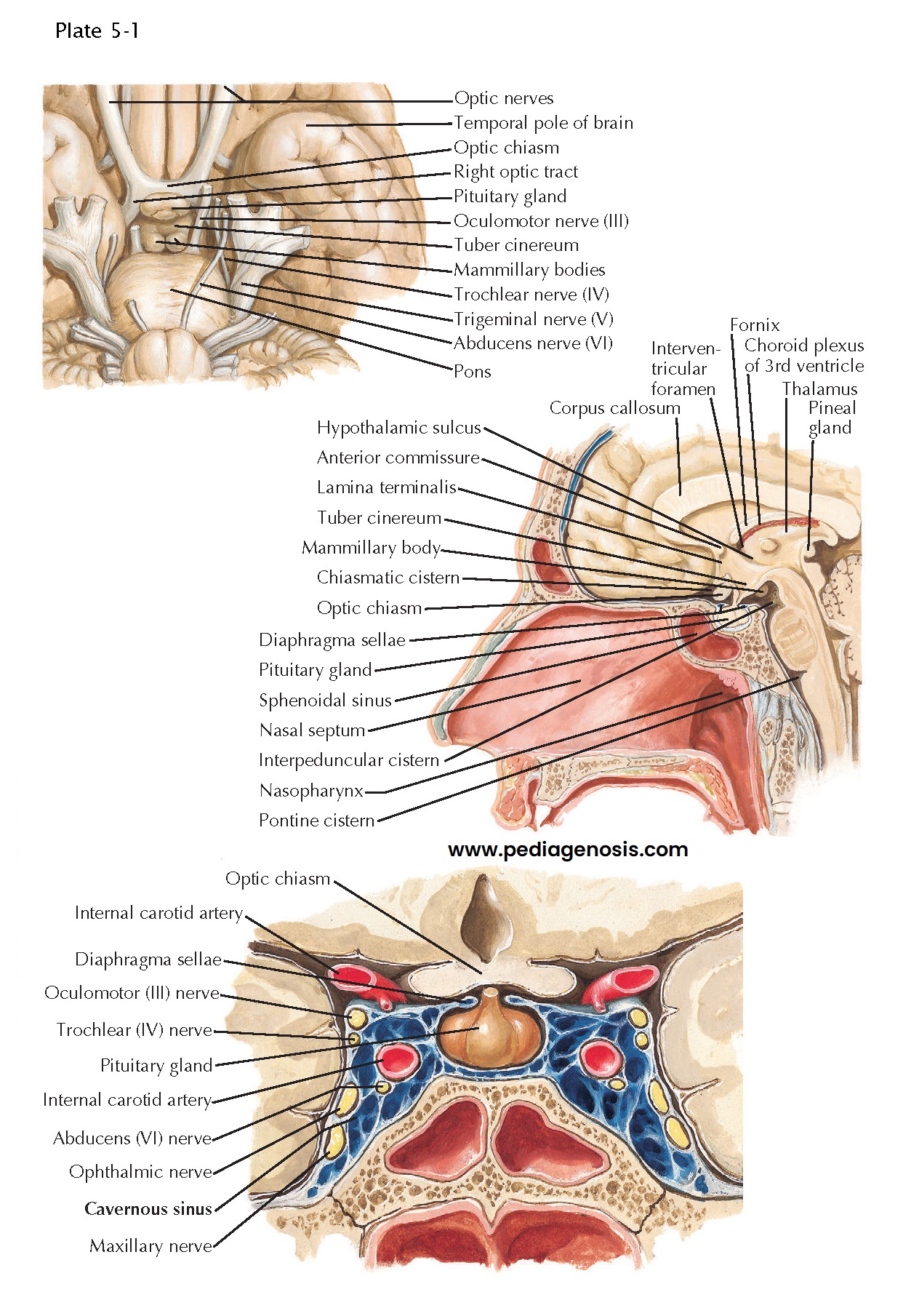
Perhaps for this reason, the hypothalamus is particularly well protected. It lies at the base of the skull, just above the pituitary gland, to which it is attached by the infundibulum, or pituitary stalk. As a result, trauma that affects the hypothalamus would almost always be lethal. It receives its blood supply directly from the circle of Willis (see Plate 5-3), so it is rarely compromised by stroke, and it is bilaterally reduplicated, with survival of either side being sufficient to sustain normal life.
On the other
hand, the hypothalamus may be involved by a number of pathologic processes that
arise from structures that surround it, and the signs and symptoms that first
attract attention in those disorders are often due to the involvement of those
neighboring structures. Examination of the ventral surface of the brain shows
that the hypothalamus is framed by fiber tracts. The optic chiasm marks the
rostral extent of the hypothalamus, and the optic tracts and cerebral peduncles
identify its lateral borders. The pituitary stalk emerges from the midportion
of the hypothalamus, sometimes called the tuber cinereum (gray swelling), just
caudal to the optic chiasm. As a result, tumors of the pituitary gland, which
are among the more common causes of hypothalamic dysfunction, typically involve
the optic chiasm (producing bitemporal visual field defects) or the optic
tracts as an early sign.
The posterior
part of the hypothalamus is defined by the mammillary bodies, which are
bordered caudally by the interpeduncular cistern, from which emerge the
oculomotor nerves. These are joined in the cavernous sinus, which runs just
below the hypothalamus and lateral to the pituitary gland, by the trochlear and
abducens nerves. Hence pathologies such as aneurysms of the internal carotid
artery or infection or thrombosis of the cavernous sinus, which may impinge on
the hypo- thalamus, typically involve the nerves controlling eye movements at
an early stage. If there is a mass of suf- ficient size, it may also involve
the trigeminal nerve. The ophthalmic division, which traverses the cavernous
sinus, is most commonly involved, but if the mass is large enough and
posteriorly located, it can involve the maxillary or even the mandibular
division of the trigeminal nerve as well. Just lateral to the cavernous sinus
sits the medial temporal lobe. As a result, pathology in this area can also
cause seizures, most commonly of the complex partial type, with loss of
awareness for a brief period.
In the midline, the hypothalamus borders the ventral part of the third ventricle. The supraoptic recess of the third ventricle, which surmounts the optic chiasm, ends at the lamina terminalis, the anterior wall of the ventricle. This is the most anterior part of the diencephalon in the developing brain. The infundibular recess defines the floor of the hypothalamus that overlies the pituitary stalk. This portion of the hypothalamus is called the median eminence and is the site at which hypothalamic releasing hormones are secreted into the pituitary portal circulation (see Plate 5-3).


