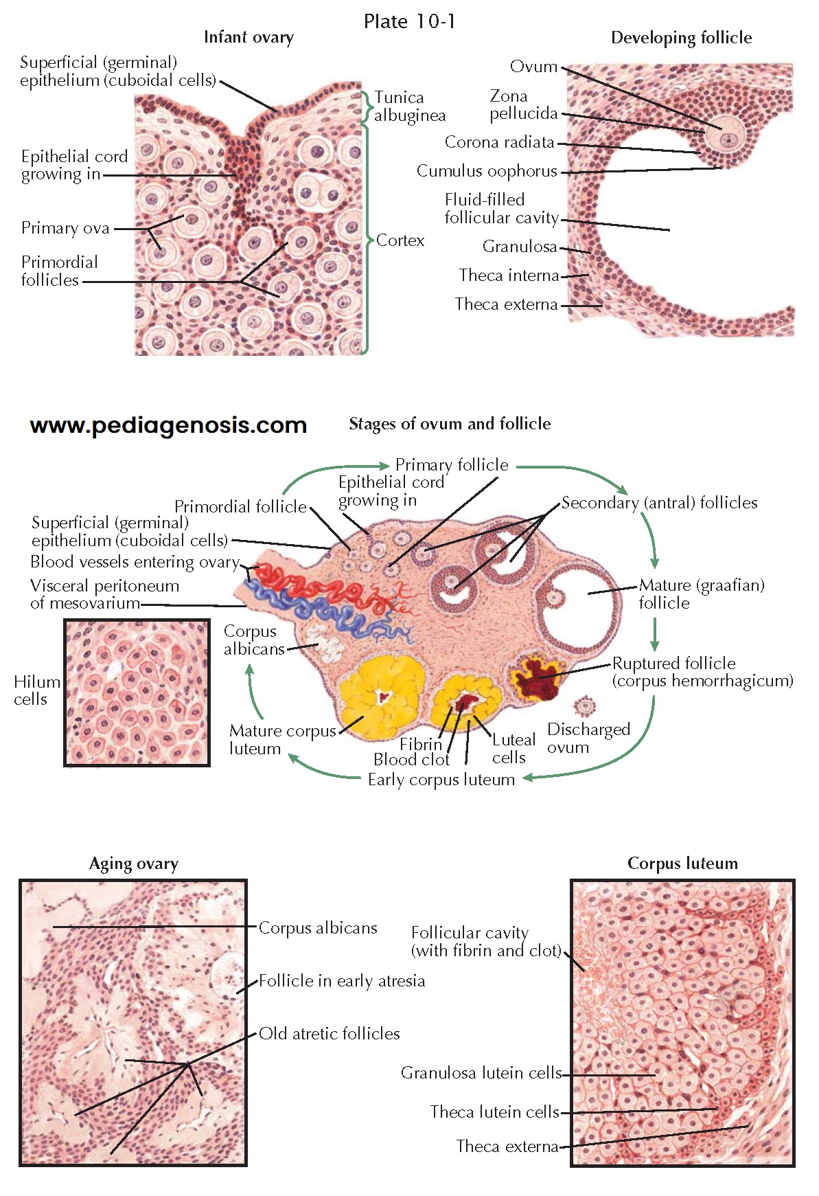
OVARIAN STRUCTURES AND DEVELOPMENT
The ovaries develop
from a thickening of cells that form ridges medial to the müllerian and wolffian
bodies. These germinal ridges appear at the sixth week. Primary oocytes,
arising in the umbilical vesicle (yolk sac) and migrating along the mesentery
of the hindgut, arrive in the embryonic gonads and are thought to provide the
countless thousands of ova that crowd the ovary at birth.
By the third month of gestation, the ovaries descend toward the pelvis. The pull of the gubernaculum—an abdominal fold that grows more slowly than the rest of the fetus—exerts a downward traction on the gonadal ridges. Later, these folds fuse in their midportion with that part of each müllerian duct that develops into the uterine fundus. The lateral half and the medial portion of the folds become the round ligaments and the suspensory ligaments of the ovary, respectively.
The infant ovary is a sausage-shaped structure, with a pale and smooth
surface. A gradual thickening and shortening occur throughout the first decade
of life. The major gain in size and weight takes place after the menarche and
during adolescence. Two layers, the germinal epithelium and the tunica
albuginea, constitute the surface of the prepubertal ovary. They are crowded
with primordial ova that are surrounded by dark-staining cells, the origin of
the future granulosa cells. The granulosa cells are polygonal and rather
uniform, round, with sharply outlined nuclei in a poorly stainable cytoplasm
that contains, however, numerous granules, from which this layer derives its
name.
As the primordial follicle develops, it sinks, with its single layer of
epithelial cells, toward the center of the ovary. The attendant cells
proliferate to form a manylayered coating of granulosa cells around the
developing follicle. A crescentic cavity forms eccentrically, in which
follicular fluid accumulates. From the surrounding ovarian stroma, a capsule of
theca cells differentiates. The theca interna is rich in capillaries, upon
which the avascular theca granulosa must depend for nourishment. Before
menarche, while still little or no follicle-stimulating hormone is present,
these follicles develop no further but degenerate and become atretic.
The mature gonad is an approximately almond-shaped structure, pitted and
scarred by the stigmata of ovulation. Spiral arteries enter at the hilum and
are involved in sequential changes during the cyclic ebb and sway of follicle
growth and development of corpora lutea. In the hilum are also found cells with
morphologic and histochemical properties, similar to the interstitial cells of
the testis—vestiges from the fetal period, before sex differentiation took
place. Proliferation of these cells or tumor formation may result in
virilization.
In the ripening follicle, the oocyte is a spherical body composed of
clear protoplasm. It contains a round, dark-staining nucleus, with a definite
surrounding membrane and an eccentric nucleolus. A transparent membrane, the
zona pellucida, encloses a fluid-filled perivitelline space in which the egg
floats freely. A dense layer of granulosa cells, the cumulus oophorus, closely
envelops the egg and attaches it to the follicular wall. The cumulus cells
immediately next to the zona arrange themselves radially outward to form the
corona radiata.
The two-layered theca envelope coats the follicle. The theca interna is
composed of large epithelioid cells interspersed in connective tissue and rich
in blood and lymph vessels. The theca externa is thick and dense, consisting of
circularly arranged connective tissue fibers.
In the follicles that do not mature but degenerate, the granulosa layer first becomes disorganized. The corona loses its radial arrangement. Thereafter, the follicular cavity shrinks, and soon the egg itself loses its characteristic features. Hyaline is deposited in a wavy, concentric band. Up to this point, the theca interna has continued to be a prominent layer of large, vesicular, nucleated cells. Degenerative changes rapidly progress until nothing is left except an amorphous hyaline scar.


