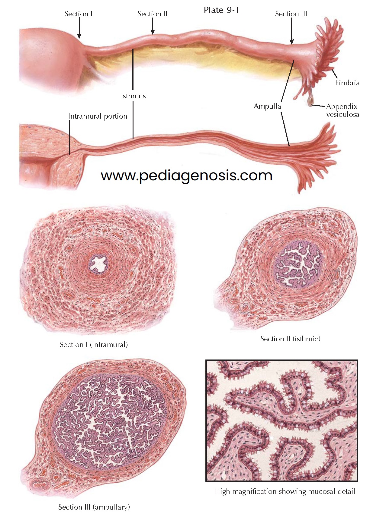
FALLOPIAN TUBES
The fallopian tubes are musculomembranous
structures, each about 12 cm in length, commonly divided into the intramural, isthmic,
and ampullary sections.
The intramural (interstitial) portion traverses the uterine wall in a more or less straight fashion. It has an ampulla-like dilation just before it communicates with the uterine cavity. On hysterosalpingography, this tiny tubal antrum either is connected with the shadow of the uterine cavity by a threadlike communication or is separated from it by a narrow, empty zone. This constric- tion of the tubal shadow, usually designated as the tubal sphincter, is caused by an annular fold of the uterine mucosa at the junction of both organs.
The isthmic portion is not quite so narrow as the intramural part. Its course
is slightly wavy. The ampullary portion is tortuous and gradually widens toward
its outer end. It terminates in a fimbriated infundibulum, which resembles a ruffled
petunia or sea anemone. One of the fimbriae, the fimbria ovarica, is grooved and runs
along the lateral border of the mesosalpinx to the ovary. Frequently, one or more
small vesicles filled with clear, serous fluid, called appendices vesiculosae or hydatids
of Morgagni, are attached to the fringes of the tube by a thin pedicle. They are
remnants of mesonephric tubules.
The wall of the tube consists of three layers—a serosal coat, a muscular layer,
and a mucosal lining. The tunica muscularis is composed of an inner circular and
an outer longitudinal layer of smooth muscle fibers. The interstitial portion is
equipped with an additional, innermost, longitudinal muscle layer. The muscular
coat is thicker in the medial section than in the ampullary portion, where the longitudinal
muscle bundles are more widely separated. Contraction of the longitudinal
muscle fibers of the ovarian fimbria brings the infundibulum in close contact with
the surface of the ovary. The abundant blood supply of the tubes is derived
from the ovarian and uterine blood vessels. The blood vessels are strikingly abundant,
particularly in the infundibulum and the fimbriae, where they form with
interspersed muscle bundles a kind of erectile tissue, which, if engorged, enables
the tube to sweep over the surface of the ovary.
The mucosa (endosalpinx) is thrown into longitudinal folds that are sparse,
low, and broad in the inner portions but numerous, branched, and slender in the
ampullary portion. The simple mucosal arrangement of the inner sections contrasts
strikingly with the complicated, labyrinth-like appearance of the arborescent
mucosa in the ampulla.
The mucosa rests on a thin basement membrane, is connected with the muscularis
by a thin layer of loose, vascular connective tissue, and can undergo moderate decidual
reaction if a fertilized ovum becomes implanted in the tube.
The mucosa consists of a single layer of columnar cells, some of which are
ciliated, whereas others are secretory. A third group of peg-shaped cells with dark
nuclei is intercalated between the ciliated and secretory cells and represents apparently
worn-out cells, which are gradually cast out into the lumen of the tube. The
numerical relationship between ciliated and secretory cells changes during the menstrual
cycle. The number of secretory cells that secrete nutrient material to sustain the
ovum during its stay in the tube is greater in the luteal phase. The height of the
tubal epithelium reaches its peak during the period of ovulation and is lowest during
menstruation.
The motion of the tubal cilia is directed toward the uterus, causing a current that can sweep small particles into the uterus, but peristaltic action and no the ciliary motion drives the ovum toward the uterus.


