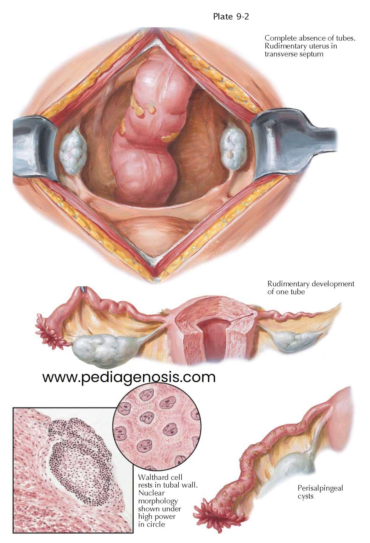
CONGENITAL ANOMALIES I—ABSENCE, RUDIMENTS
The fallopian tubes arise from the unfused cranial portions of the müllerian (paramesonephric) ducts. The origin of these ducts is first indicated in embryos of 8.5 to 10 mm by grooves in the thickened epithelium of the urogenital ridge lateral to the wolffian ducts, which were formed in an earlier stage of development. Soon these grooves become deeper and advance caudad as tubes until they meet the epithelium of the urogenital sinus in a prominence, the müllerian tubercle.
The development and growth of the müllerian ducts depend on the presence of
the wolffian ducts. As a rule, the müllerian duct fails to develop when the ipsilateral
wolffian duct is lacking. In the absence of müllerian inhibiting factor made by the
Sertoli cells of the developing testes, the mesonephric ducts regress, and the
paramesonephric ducts develop into the female genital tract. The more cephalad portions
of the paramesonephric ducts, which open directly into the peritoneal cavity,
form the fallopian tubes.
The paramesonephric ducts, and similarly the wolffian duct, grow more slowly
than the fetal trunk as a whole. As a result, the location of the coelomic ostia
of the ducts gradually slides caudally to the fourth lumbar segment. Originally,
the urogenital fold, which contains the mesonephros and the wolffian and müllerian
ducts, has a sagittal course parallel to the spine. The development and growth of
the adrenals and kidneys cephalad to these structures change the direction of the
urogenital fold, bending it laterally in its upper section. With progressive descent
of the ovary and the fallopian tube, the direction of the tube becomes more transverse,
whereas the lower parts of the müllerian ducts, which give rise to the uterus, retain
their original longitudinal course and fuse in the midline.
The muscular and connective tissues of the tubes are first apparent in the
third month of fetal life and may be clearly demonstrated in the fifth month. However,
the ampullary portion does not take on its characteristic appearance until the last
month of gestation.
Congenital anomalies of the fallopian tubes may be due to developmental error
or injury, such as torsion or inflammation. Complete absence of both tubes is sometimes
observed in combination with aplasia of the uterus. In such cases, a transverse
pelvic septum, corresponding to the broad ligament, connects the ovaries and may
contain muscular nodules in the lateral sections of its upper margin that represent
rudiments of the müllerian ducts. The round ligaments, which have a separate
origin, may be well developed. In these cases aplasia of the vagina is common but
not the rule.
Absence of the ampullary portion of the tube is probably caused by twisting
and subsequent necrosis with resorption of the ampullary portion of the tube rather
than a result of developmental failure. A congenital defect of the wolffian duct
is, as a rule, followed by a defect of the homolateral müllerian duct. The resulting
anomaly is characterized by the absence of the fallopian tube, the uterine horn,
the kidney, and the ureter.
Small, solid, or cystic nodules of a type first described by Walthard and thus
called “Walthard cell rests” are commonly found in the subserosal tissue of the
fallopian tube. The nuclei of these Walthard cells show a median groove or fold,
which is a fundamental characteristic of these cells.
Frequently found on the surface of otherwise normal fallopian tubes are tiny multiple serosal (perisalpingeal) cysts. They apparently originate from an invagination and occlusion of the serosal epithelium and do not have any practical significance. The supposition of some authors that these cysts are caused by chronic inflammation is not generally accepted.


