Valves
 |
| CARDIAC VALVES OPEN AND CLOSED |
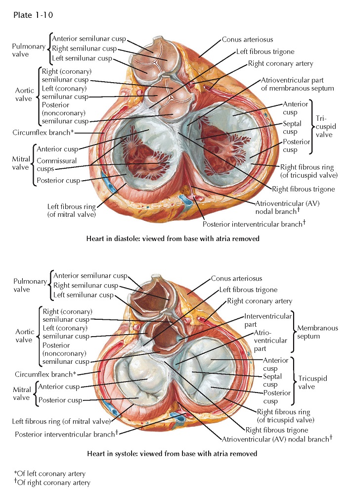
Each atrioventricular (AV) valve apparatus consists of a number of cusps, chordae tendineae, and papillary muscles. The cusps are thin, yellowish white, glistening trapezoid-shaped membranes with fine, irregular edges. They originate from the annulus fibrosus, a poorly defined and unimpressive fibrous ring around each AV orifice. The amount of fibrous tissue increases only at the right and left fibrous trigones.
The atrial surface of the AV valve
is rather smooth (except near the free edge) and not well demarcated from the
atrial wall. The ventricular surface is irregular because of the insertion of
the chordae tendineae and is separated from the ventricular wall by a narrow
space. The extreme edges of the cusps are thin and delicate with a sawtooth
appearance from the insertion of equally fine chordae. Away from the edge, the
atrial surface of the cusps is finely nodular, particularly in small children.
These nodules are called the noduli Albini. On closure of an AV valve, the
narrow border between the row of Albini nodules and the free edge of each cusp
presses against that of the next, resulting in a secure, watertight closure.
The chordae tendineae may be divided into the
following three groups:
The chordae of the first order
insert into the extreme edge of the valve by a large number of very fine
strands. Their function seems to be merely to prevent the opposing borders of
the cusps from inverting.
The chordae of the second order
insert on the ventricular surface of the cusps, approximately at the level of
the Albini nodules, or even higher. These are stronger and less numerous. They
function as the mainstays of the valves and are comparable
to the stays of an umbrella.
The chordae of the third order
originate from the ventricular wall much nearer the origin of the cusps. These
chordae often form bands or foldlike structures that may contain muscle.
The first two groups originate from
or near the apices of the papillary muscles. They form a few strong, tendinous
cords that subdivide into several thinner strands as
they approach the valve edges. Occasionally, particularly on the left side, the
chordae of the first two orders may be wholly muscular, even in normal hearts,
so that the papillary muscle seems to insert directly into the cusp. This is
not surprising because the papillary muscles, the chordae tendineae,
and most of the cusps are derived from the embryonic ventricular
trabeculae and therefore were all muscular at one time.
The tricuspid valve consists
of an anterior, a medial (septal), and one or two posterior cusps.
The depth of the commissures between the cusps is variable, but the
commissures never reach the annulus, so the cusps are only incompletely
separated from each other.
 |
| VALVES AND FIBROUS SKELETON OF HEART |
The mitral (bicuspid) valve
actually is made up of four cusps: two large ones—the anterior (aortic)
and posterior (mural) cusps—and two small commissural cusps.
Here, as in the tricuspid valve, the commissures are never complete, and they
should not be so constructed in the surgical treatment of mitral stenosis.
The arterial or semilunar valves
differ greatly in structure from the AV valves. Each consists of three
pocketlike cusps of approximately equal size. Although, functionally the
transition between the ventricle and the artery is abrupt and easily
determined, this cannot be done anatomically in any simple manner. There is no
distinct, circular ring of fibrous tissue at the base of the arteries from which these and the valve
cusps arise; rather, the arterial wall expands into three dilated pouches, the
sinuses of Valsalva, whose walls are much thinner than those of the aorta or
pulmonary artery. The origin of the valve cusps is therefore not straight but
scalloped.
The cusps of the arterial semilunar
valve are largely smooth and thin. At the center of the free margin of each
cusp is a small fibrous nodule called the nodulus
Arantii. On each side of the nodules of Arantius, along
the entire free edge of the cusp, there is a thin, half-moon–shaped area
called the lunula that has fine striations parallel to the edge. The
lunulae are usually perforated near the insertion of the cusps on the aortic
wall. In valve closure, because the areas of adjacent lunulae appose each
other, such perforations do not cause insufficiency of the valve and are
functionally of no significance.


