Suprahyoid Muscles Anatomy
1.
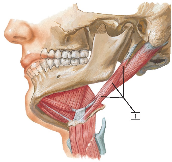
Stylohyoid muscle
Origin:
Arises
from the styloid process of the temporal bone.
Insertion:
Attaches
to the body of the hyoid bone.
Action:
Elevates
and retracts the hyoid bone in an action that elongates the floor of the mouth.
Innervation: Facial nerve.
Comment:
The
stylohyoid muscle is perforated near its insertion by the tendon of the 2
bellies of the digastric muscle.
The
stylohyoid is 1 of the 3 muscles arising from the styloid process, each innervated
by a different cranial nerve. The other 2 muscles are the
stylopharyngeus (CN IX) and the styloglossus (CN XII).
Clinical:
The
stylohyoid is one of several muscles that help stabilize the
hyoid bone, which is important in movements of the tongue and in swallowing. If
this process is compromised, these movements become
more difficult and/or painful to execute.
1.
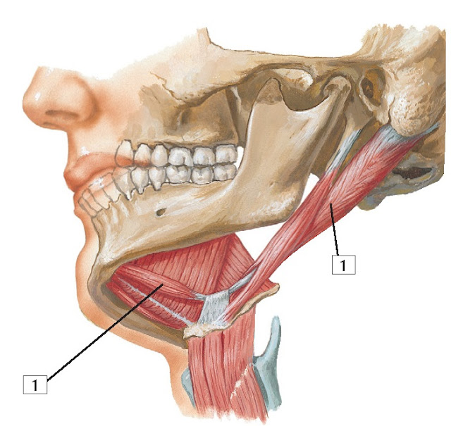
Digastric muscle
Origin:
The
digastric muscle consists of 2 bellies. The posterior belly is the longest, and
it arises from the mastoid notch of the temporal bone. The anterior belly
arises from the digastric fossa of the mandible.
Insertion:
The
2 bellies end in an intermediate tendon that perforates the stylohyoid muscle
and is connected to the body and greater horn of the hyoid bone.
Action:
Elevates
the hyoid bone and, when both muscles act together, helps the lateral pterygoid
muscles open the mouth by depressing the mandible.
Innervation:
The
anterior belly is innervated by the mylohyoid nerve, a branch of the mandibular
division of the trigeminal nerve. The posterior belly is innervated by the
facial nerve.
Comment:
The
2 bellies of the digastric muscle are unique because they
are innervated by different cranial nerves.
Clinical:
The
digastric muscles are important for opening the mouth symmetrically and are
assisted by the lateral pterygoid muscles.


