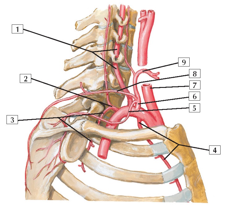
Subclavian Artery Anatomy
1. Vertebral artery
2. Costocervical trunk
3. Supreme intercostal artery
4. Internal thoracic artery
5. Suprascapular artery
6. Thyrocervical trunk
7. Common carotid artery
8. Transverse cervical artery
9. Inferior thyroid artery
Comment: The subclavian artery is divided into 3 parts
relative to the anterior scalene muscle. The 1st part is medial to the muscle,
the 2nd is posterior, and the 3rd is lateral. Branches of the subclavian
include the vertebral and internal thoracic (mammary) arteries, thyrocervical
and costocervical trunks, and dorsal scapular artery.
The vertebral artery ascends through
the C6-1 transverse foramina and enters the foramen magnum. The internal
thoracic descends parasternally. The thyrocervical trunk supplies the thyroid
gland (inferior thyroid), the lower region of the neck (transverse cervical),
and the dorsal scapular region (suprascapular). The costocervical trunk
supplies the deep neck (deep cervical) and several intercostal spaces (supreme
intercostal). The dorsal scapular branch is inconstant;
it may arise from the transverse cervical artery.
Clinical: The branches of the subclavian artery anastomose
with branches of the axillary artery around the shoulder joint, with branches
of the thoracic aorta (intercostal branches) along the rib cage, across the midline of the neck and face via
branches from both external carotid arteries, and with the internal carotid
arteries and the vertebral branches (circle of Willis on the brainstem). These
interconnections are important if the vasculature in
1 region is compromised.


