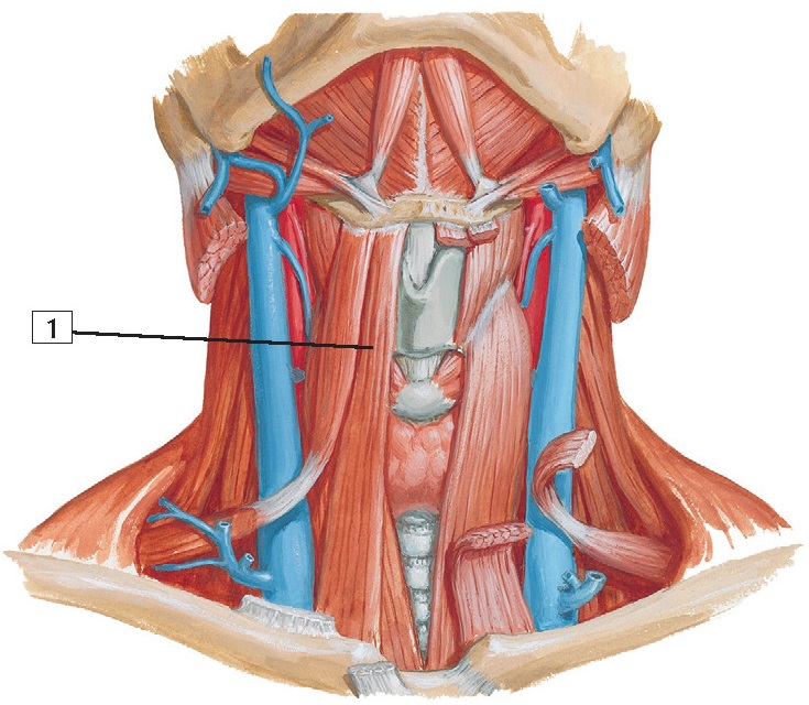
Infrahyoid and Suprahyoid Muscles Anatomy
1.
Sternohyoid muscle
Origin:
Manubrium
of the sternum and medial portion of the clavicle.
Insertion:
Body
of the hyoid bone.
Action:
Depresses
the hyoid bone after swallowing.
Innervation: C1, C2, and C3 from the ansa cervicalis.
Comment:
The
sternohyoid is part of the group of infrahyoid muscles. These muscles are often
referred to as “strap” muscles because they are long and narrow.
Clinical:
The
infrahyoid, or “strap,” muscles are surrounded by an investing layer of
cervical fascia that binds the neck muscles in a tight fascial sleeve. Swelling
within this confined space can be painful and potentially damaging to adjacent
structures.
Immediately
deep to this investing fascia is a “pretracheal space” anterior to the trachea
and thyroid gland, which can provide a vertical conduit
for the spread of infections.
1.
Sternothyroid muscle
Origin:
Arises
from the posterior surface of the manubrium of the sternum.
Insertion:
Attaches
to the oblique line of the thyroid cartilage.
Action:
Depresses
the larynx after the larynx has been elevated for swallowing.
Innervation:
C2
and C3 from the ansa cervicalis.
Comment:
The
sternothyroid is part of the group of infrahyoid muscles. Because they are long
and narrow, these muscles are often referred to as
“strap” muscles.
Clinical:
The
infrahyoid, or “strap,” muscles are surrounded by an investing layer of
cervical fascia that binds the neck muscles in a tight fascial sleeve. Swelling
within this confined space can be painful and potentially damaging to adjacent
structures.
Immediately
deep to this investing fascia is a “pretracheal space” anterior to the trachea
and thyroid gland, which can provide a vertical conduit
for the spread of infections.
1.
Omohyoid muscle
Origin:
This
muscle consists of an inferior and a superior belly. The inferior belly arises
from the superior border of the scapula, near the suprascapular notch.
Insertion:
The
muscle is attached by a fibrous expansion to the clavicle and forms the
superior belly, which inserts into the inferior border of the hyoid bone.
Action:
Depresses
the hyoid bone after the bone has been elevated. Also retracts and steadies the
hyoid bone.
Innervation:
C1,
C2, and C3 by a branch of the ansa cervicalis.
Comment:
The
omohyoid acts with the other infrahyoid muscles to depress the larynx and hyoid
bone after these structures have been elevated during swallowing.
The
omohyoid is an unusual “strap” muscle because it arises from the
scapula in the shoulder region.
Clinical:
The
infrahyoid, or “strap,” muscles are surrounded by an investing layer of
cervical fascia that binds the neck muscles in a tight fascial sleeve. Swelling
within this confined space can be painful and potentially damaging to adjacent
structures.
Immediately
deep to this investing fascia is a “pretracheal space” anterior to the trachea
and thyroid gland, which can provide a vertical conduit
for the spread of infections.
1.
Thyrohyoid muscle
Origin:
Arises
from the oblique line of the lamina of the thyroid cartilage.
Insertion:
Attaches
to the inferior border of the body and the greater horn of the hyoid bone.
Action:
Depresses
the hyoid bone and, if the hyoid bone is fixed, draws the thyroid cartilage
superiorly.
Innervation:
C1
via the hypoglossal nerve (CN XII).
Comment:
The
thyrohyoid muscle is supplied by fibers of the 1st cervical nerve that happen
to travel with the last cranial, or hypoglossal, nerve (CN XII).
The
thyrohyoid muscle is also one of the infrahyoid, or “strap,” muscles.
Clinical:
Trauma
to the neck may damage the ansa cervicalis (C1-3) and its branches, leading to
paralysis of the infrahyoid and suprahyoid muscles. Because these muscles are
critical in the process of swallowing, dysphagia (difficulty in swallowing) may ensue.


