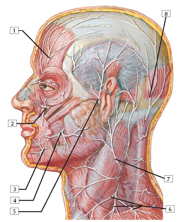
Cutaneous Nerves of Head
and Neck Anatomy
1. Supra-orbital nerve
2. Infra-orbital nerve
3. Mental nerve
4. Buccal nerve
5. Auriculotemporal nerve
6. Supraclavicular nerves (C3, C4)
7. Great auricular nerve (C2, C3)
8. Greater occipital nerve (C2)
Comment: Cutaneous innervation of the face is by the 3
divisions of the trigeminal nerve (CN V). The ophthalmic division is
represented largely by the supra-orbital and supratrochlear nerves. The
maxillary division is represented by the infra-orbital and zygomaticotemporal
nerves. The mandibular division is represented largely by the mental, buccal,
and auriculotemporal nerves.
The skin on the back of the scalp
receives cutaneous innervation from the greater occipital nerve (dorsal ramus
of C2); the skin on the back of the neck receives innervation from dorsal rami
of cervical nerves.
The 1st cervical nerve (C1) has few if
any sensory nerve fibers from the skin, so it
is usually not shown on dermatome charts.
Clinical: The sensory innervation of the face is via the 3 divisions of CN V. Trauma anywhere along the
pathway of the nerve, including that on the face itself (e.g., facial
lacerations), can lead to loss of sensation. The innervation of the muscles of
facial expression will not be affected unless a laceration also damages the terminal branches of the facial nerve.


