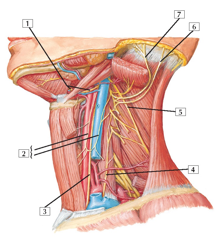
Cervical Plexus In Situ Anatomy
1. Hypoglossal nerve (CN XII)
2. Ansa cervicalis (Superior root;
Inferior root)
3. Vagus nerve (CN X)
4. Phrenic nerve
5. Accessory nerve (CN XI)
6. Lesser occipital nerve
7. Great auricular nerve
Comment: The cervical plexus arises from ventral rami of
C1-4. It provides motor innervation for many of the muscles of the anterior and
lateral compartments of the neck. This plexus also provides cutaneous
innervation to the skin of the neck.
Most of the motor contributions to the
infrahyoid muscles arise from a nerve loop called the ansa cervicalis (C1-3).
The cervical plexus also gives rise to
the first 2 of 3 roots contributing to the phrenic nerve (C3, C4, and C5). The
phrenic nerve innervates the abdominal diaphragm.
Clinical: Unilateral trauma to the posterior cervical
triangle of the neck may injure the accessory nerve (CN XI) (ipsilateral
innervation of the sternocleidomastoid and trapezius muscles), the phrenic
nerve (C3-5) (innervates the ipsilateral hemi- diaphragm), or the trunks or cords
of the brachial plexus. The integrity of each of these nerves should be
assessed when trauma is evident.


