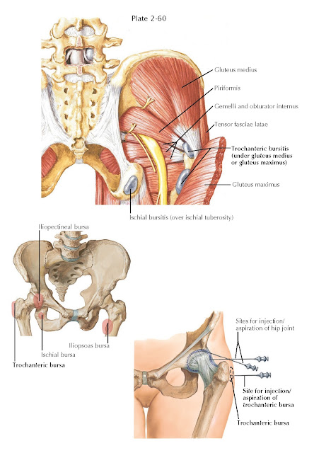
TROCHANTERIC BURSITIS
Lateral (or trochanteric) hip pain is a very common presenting complaint. Patients often report the insidious onset of pain over the hip bone (trochanter). Occasionally, this can be caused by trauma or direct injury to the prominence. Complaints include activity-related increases in pain as well as occasionally pain at rest. Often there will be difficulty lying on the affected side for sleep. Pain is usually described as sharp and burning. It may radiate down the lateral aspect of the thigh along the course of the iliotibial band.
The true cause of greater trochanteric pain syndrome is often elusive.
Onset can at times be due to a specific traumatic event. However, this is less
often the situation. Trochanteric hip pain may be referred from the hip joint
itself owing to osteoarthritis or a labral pathologic process. Proximal femoral
anatomy (excessive proximal femoral varus) and hip abductor weakness are
thought to predispose patients to trochanteric bursitis and pain.
A general hip examination is performed as discussed previously, with the
focus on hip range of motion and hip abduction strength (Trendelenburg test),
as well as a gait examination. Palpation of the lateral hip should help guide
the cause of pain. Palpation must take place over the greater trochanter,
gluteus medius tendon, and piriformis, because they all may contribute to this
syndrome. The Ober test is also performed to determine if the iliotibial band
is contracted.
Anteroposterior pelvis, true anteroposterior, and lateral views of the
affected hip are standard. MRI of the hip is indicated with pain recalcitrant
to treatment modalities when minimal osteoarthritis is seen on plain
radiographs.
The differential diagnosis includes trochanteric bursitis, hip abductor
tear/tendonitis, piriformis syndrome, hip osteoarthritis, hip osteoarthritis,
and lumbar disc disease/spondylosis/sacroiliitis.
Initial treatment of trochanteric bursitis includes correcting any
observable abductor weakness or iliotibial band contracture with physical
therapy. NSAIDs are also used for pain relief. Injection may be considered at
any point. If severe pain is present on initial presentation
with negative radiographs, corticosteroid injection may help alleviate the symptoms
and allow the proper rehabilitation.
Persistent trochanteric tenderness often necessitates advanced imaging.
MRI is used to evaluate the state of the abductor muscle complex. Partial
tearing or complete rupture may be present. In this scenario, greater
trochanteric pain syndrome recalcitrant to physical
therapy and NSAIDs may require shock-wave therapy or injection of platelet-rich
plasma into the tendinotic/partially torn area. If a labral or intra-articular
pathologic process is suspected, an arthrogram may be performed at the time of
the MRI with local anesthetic to determine if the pain may be referred from the
joint. Treatment at that point would be guided by results of both the
arthrographic and imaging findings.


