PHYSICAL EXAMINATION
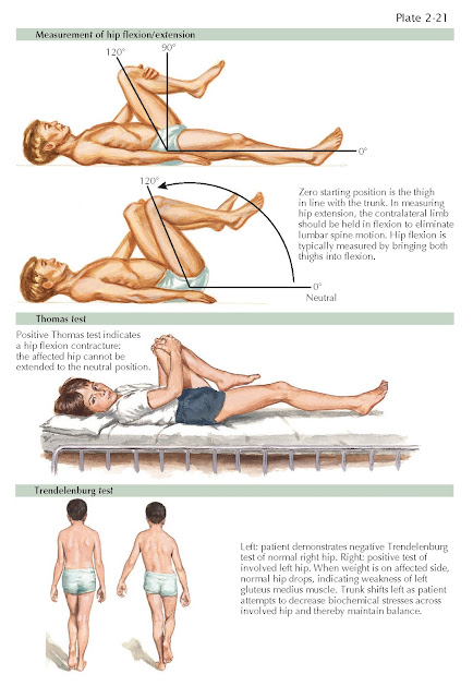
Physical examination of the hip is initiated with observation of the patient. Specific note is made of body habitus. Gait is evaluated directly, looking for a Trendelenburg or antalgic gait that favors the affected side. Both gait patterns are associated with intra-articular and extra-articular hip pathologic processes.
The patient is then placed supine on an examination table, and landmarks
are palpated. These landmarks include the anterior superior iliac spine, iliac
crest, pubic symphysis, pubic tubercles, and proximal adductor tendons. The
abdomen may also be palpated in this position if there is concern about any
abdominal disorder.
Range of motion of the hips is then evaluated. This may be also examined
combined with the upright and prone position based on the preference of the
examiner. First checked is hip flexion, followed by internal and external
rotation with the hip flexed to 90 degrees. In full extension, the hip is taken
into adduction as well as abduction. Straight-leg hip flexion can also be evaluated
for hamstring tightness or radicular low back pain. Deficits or painful ranges
are noted compared with the contralateral/unaffected side. Specific tests for
the hip joint in this position include the anterior impingement test
(flexion/adduction/internal rotation), which is commonly painful in the face of
labral tears or osteoarthritis. The hip may also to be placed in the figure-4
position, looking for pain or a loss of motion that may also indicate
intra-articular findings. Straight-leg raising is performed to evaluate hip
flexion strength in the supine position. Resisted adduction is also tested in
the supine position to evaluate strength and integrity of the adductors.
The patient is then placed in the lateral decubitus position, and both
the affected and nonpainful side are evaluated. Palpation is begun over the
greater trochanter (trochanteric bursa posterior/superiorly), the gluteus
medius/minimus tendons just proximal to the tip of the trochanter, and the
piriformis tendon at the proximal/posterior aspect of the trochanter just
posterior to the abductor musculature. The iliotibial band is checked for
tightness with the Ober test. Hip abduction strength is evaluated with the hip
held in the abducted position against resistance.
A prone examination is then completed. Examination of the hamstrings is
best completed in this position, as well as palpation of the ischial tuberosity
and proximal hamstring insertion and muscle bellies. Hamstring
strength is tested with resisted knee flexion at 90 and 45 degrees.
The Thomas test is useful to look for hip flexion contracture while the patient
is in this position.
Completion of the hip examination is then done in the upright position.
Palpation of the posterior iliac crest and posterior superior iliac
spine/sacroiliac joint is completed at this point. This position also allows
for palpation of the midline and paraspinal lumbar spine. Hip flexion strength
in this position isolates the iliopsoas unit.
The joint proximal (lumbar spine) and distal (knee) are also evaluated
for completeness and to rule out referred pain. This may also be inferred from
good history taking.


