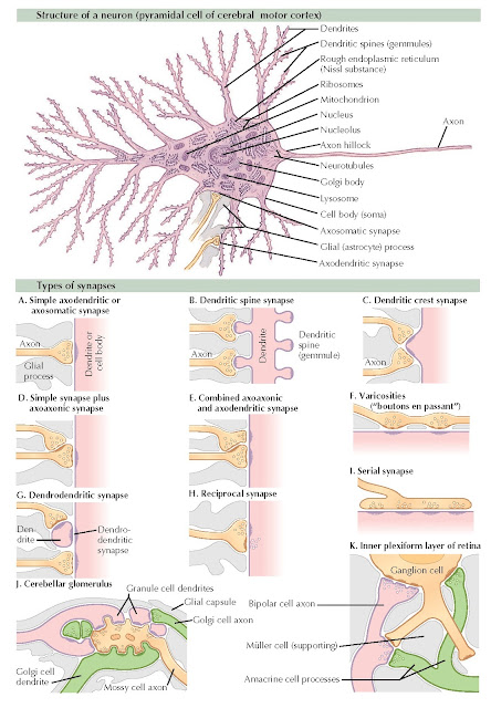
NEURONAL STRUCTURE AND SYNAPSES
A typical neuron of the central nervous system consists of three parts: dendritic tree, cell body (soma), and axon. The highly branched dendritic tree has a much greater surface area than the remainder of the neuron and is the receptive part of the cell. Incoming synaptic terminals make contact directly with the dendritic surface or with the small spines (gemmules) that protrude from it. The membrane potential induced in the dendrites spreads passively onto the cell soma, which allows all inputs acting on the neuron to summate in controlling the rate of neuronal discharge through the axon.
The soma contains the various
organelles that control and maintain neuronal structure: nucleus, Golgi
apparatus, lysosomes, ribosomes, mitochondria, and smooth and rough endoplasmic
reticula. The rough endoplasmic reticulum, studded with ribosomes, is called
the Nissl substance because of its characteristic blue staining with
Nissl stain. The ribosomes are the site of synthesis of neuronal
proteins; as in other cells, the ribonucleic acid (RNA) templates that control
protein structure are transcribed from patterns in the nuclear deoxyribonucleic
acid (DNA). The soma membrane is also covered with synaptic endings separated
by glial processes. Because of their proximity to the origin of the axon, these
synaptic endings have an especially potent effect on the rate of discharge of
the neuron.
 |
| Plate 2-14 |
In humans, the axon can
extend for several feet. Such lengths pose supply problems because the neuron
must transport proteins and other synthesized substances as far as the axon
terminals. Certain key substances are transported, at a rate as high as 400
mm/day, by rapid axonal transport, a process probably associated with the
microtubules that originate in the soma and run the length of the axon. Other
soluble and particulate substances move by slow axonal transport at a
rate of 1 to 4 mm/day, aided partly by the peristalsis-like motion of the axon.
The axon originates from a conical
projection (axon hillock) on the soma (as shown in Plate 2-14) or on one of the proximal dendrites. The axon membrane is
specialized for the transmission of action potential. Because of its shape and
high excitability, the initial segment of the axon is usually the site of
action potential generation. The action potential then spreads down the axon
and back to the soma and proximal dendrites. Because of the low excitability of
the dendrites, the impulse usually does not spread very far into the dendritic
tree. At its distal end, the axon divides into numerous branches, which end in
synapses.
The most common central nervous
system (CNS) synapses are those between axon terminals and dendrites
(axodendritic) or between axon terminals and somata (axosomatic). Axodendritic
synapses take several forms. Spine synapses are of particular interest,
because they may be the site of morphologic changes accompanying learning. Axosomatic
synapses are of the simple type shown in example A. Synaptic
interconnections between a number of neurons occur within structures of a
complex organization, such as the cerebellar glomerulus, although all synapses
within the glomerulus are axodendritic.
Axons also form axoaxonic
synapses with other axon terminals, and these are responsible for the
phenomenon of presynaptic inhibition. Axoaxonic synapses
are also seen in the efferent vestibular system and in connection with motor
neuron dendrites and other terminals ending on those dendrites.
The CNS also contains several less
common types of synapses. Dendrodendritic synapses are found in the
olfactory bulb. In the internal plexiform layer of the retina, synaptic
interactions involve synaptic t of bipolar, amacrine, and ganglion cell processes.
Other synapses are those formed
between the peripheral axonal processes of sensory neurons and sensory
receptor cells, as in the inner ear. Here, the axon terminal forms the
postsynaptic element that is depolarized by the presynaptic sensory cell.
There are also specialized
axosomatic synapses formed by efferent motor axons on muscle (motor end lates)
and by autonomic axons on secretory cells.


