BONES AND LIGAMENTS AT HIP
The femur is the longest and strongest bone in the body, comprising a shaft and two irregular extremities that articulate at the hip and knee joints (see Plate 2-19).
The superior extremity of the bone has a nearly spherical head mounted
on an angulated neck, and prominent trochanters provide for muscular
attachments. The head is smooth, with an articular surface that is largest
above and anteriorly; this is interrupted medially by a depression, the fovea
capitis femoris, into which attaches the capitis femoris ligament.
 |
| OSTEOLOGY OF THE FEMUR |
The neck is about 5 cm long and forms an angle with the shaft,
which varies in the normal person from 115 to 140 degrees. It is compressed
anteroposteriorly and contains a large number of prominent pits for the
entrance of blood vessels.
The greater trochanter is the bony prominence of the hip. It is
palpable 12 to 14 cm below the iliac crest, is large and square, and marks the
upper end of the shaft of the femur. Its large quadrilateral surface is divided
by an oblique ridge running from the posterosuperior to its anteroinferior
angles. In front and above the ridge there is a triangular surface (which may
be smooth) for a bursa. Below and behind the ridge the bone is also smooth. Its
posterior rounded border bounds the trochanteric fossa and continues downward
as the inter- trochanteric crest. The trochanteric fossa is a deep pit on the
internal aspect of the trochanter.
The lesser trochanter is a blunt, conical projection at the
junction of the inferior border of the neck with the shaft of the femur. The
trochanters are joined behind by the intertrochanteric crest. On the anterior
surface of the femur, the junction of the neck and shaft is also ridged. This
is the intertrochanteric line, which provides attachment for the capsule of the
hip joint across the front of the bone and continues as a spiral line, winding
backward to blend into the medial lip of the linea aspera.
The shaft of the bone is fairly uniform in caliber but broadens
slightly at its extremities. It is bowed forward and its surface is smooth,
except for the thickened ridge along its posterior surface, the linea aspera.
This is especially prominent in the middle third of the bone, where lateral and
medial lips are developed. Superiorly, the lateral lip blends with the
prominent gluteal tuberosity; an intermediate lip extends as the pectineal line
to the posterior border of the lesser trochanter; and the medial lip continues
as the spiral line. The nutrient foramen of the femur, directed upward, is
located on the linea aspera.
The inferior extremity of the femur is broadened about threefold
for the knee joint. Its surfaces, except at the sides, are articular two oblong condyles
for articulation with the tibia are separated by an intercondylar fossa and
united anteriorly by the patellar surface. The wheel-like condyles are also
curved from side to side. The intercondylar fossa is especially deep
posteriorly and is separated by a ridge from the popliteal surface of the femur
above. The medial condyle is longer than the lateral condyle. The condyles rest
on the horizontal condyles of the tibia, and the shaft of the femur inclines
downward and inward.
The epicondyles bulge above and within the curvatures of the
condyles. The medial epicondyle is the more prominent,
giving attachment to the tibial collateral ligament of the knee joint. It bears
on its upper surface a pointed projection, the adductor tubercle. The lateral
epicondyle gives rise to the fibular collateral ligament. A groove below the
epicondyle borders the articular margin.
The femur is ossified from five centers: one for the shaft, one
each for the head and inferior extremity, and one for each trochanter. The
shaft is ossified at birth; ossification extends
into the neck after birth. The center for the inferior extremity of the bone
appears during the ninth month of fetal life; that for the head appears during
the first year. The center in the greater trochanter appears during ages 3 to
5; that for the lesser trochanter appears at age 9 or 10. The epiphyses for the
head and trochanters fuse with the shaft at ages 14 to 17; those at the knee
fuse with the shaft at about age 1712.
Movements of the hip joint are flexion-extension, abduction-adduction,
and medial and lateral rotation. Circumduction is also allowed.
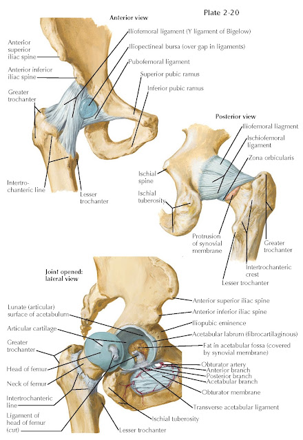
The hip joint, a synovial ball-and-socket joint, consists of the
articulation of the globular head of the femur in the cuplike acetabulum of the
coxal bone (see Plate 2-20). Compared
with the shoulder joint, it has greater stability and some decrease in freedom
of movement. The head forms about two thirds of a sphere and is covered by
articular cartilage, thickest above and thinning to an irregular line of
termination at the junction of the head and neck. The acetabulum of the coxal
bone exhibits a horseshoe-shaped articular surface arching around the
acetabular fossa. The articular fossa lodges a mass of fat covered by synovial
membrane; the transverse ligament of the acetabulum closes the fossa below. An
acetabular labrum attaches to the bony rim and to the ligament. It attaches to
the bony rim and to the ligament. Its thin, free edge cups around the head of
the femur and holds it firmly.
 |
| HIP JOINT |
The articular capsule of the joint is strong. It is attached to
the bony rim of the acetabulum above and to the transverse ligament of the
acetabulum inferiorly. On the femur, it is attached anteriorly to the
intertrochanteric line and to the junction of the neck of the femur and its trochanters.
Behind, the capsule has an arched free border, covering only two thirds of the
neck of the femur distally. Most of the fibers of the capsule are longitudinal,
running from the coxal bone to the femur, but some deeper fibers run
circularly. These zona orbicularis fibers are most marked in the posterior part
of the capsule; they help to hold the head of the femur in the acetabulum.
Three ligaments, as thickenings of the capsule, add strength. The very
strong iliofemoral ligament lies on the anterior surface of the capsule,
in the form of an inverted Y. Its stem is attached to the lower part of the
anterior inferior iliac spine, with the diverging bands attaching below to the
whole length of the intertrochanteric line. The iliofemoral ligament becomes
taut in full extension of the femur and thus helps to maintain erect posture,
because in this position the body’s weight tends to roll the pelvis backward on
the femoral heads. The pubofemoral ligament is applied to the medial and
inferior part of the capsule. Arising from the pubic part of the acetabulum and
the obturator crest of the superior ramus of the pubis, this ligament reaches
the underside of the neck of the femur and the iliofemoral ligament. The ligament
becomes taut in extension and also limits abduction. The articular capsule is
thinnest between the iliofemoral and pubofemoral ligaments but is crossed here
by the robust iliopsoas tendon. The iliopectineal bursa lies between
this tendon and the capsule. The ischiofemoral ligament forms the posterior
margin of the capsule. It arises from the ischial portion of the acetabulum and
spirals lateralward and upward, ending in the superior part of the femoral
neck. The capitis femoris ligament, about 3.5 cm long,
is intracapsular, arising from the two margins of the
acetabular notch and the lower border of the transverse acetabular ligament and
ending in the fossa of the head of the femur. It becomes taut in adduction of
the femur.
The synovial membrane of the hip joint lines the articular
capsule, covers the acetabular labrum, and is extended, sleevelike, over the
ligament of the head of the femur. The membrane covers the fat of the
acetabular notch and is reflected back along the femoral neck at the femoral
attachment of the capsule. Blood vessels to the head and neck of the femur
course under these synovial reflections.
The arteries of the hip joint are branches of the medial and
lateral circumflex femoral arteries, the deep branch of the
superior gluteal artery, and the inferior gluteal artery. The posterior branch
of the obturator artery provides a significant portion of the blood supply of
the femoral head. Nerve supply to the hip joint is derived from the
nerves supplying the quadratus femoris and rectus femoris muscles, the anterior
division of the obturator nerve (rarely also from the accessory obturator
nerve), and the superior gluteal nerve.


