VENOUS DRAINAGE OF
STOMACH AND DUODENUM
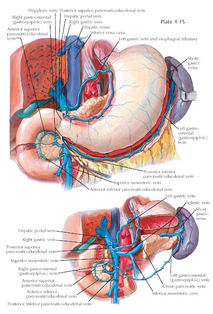
Venous blood from the stomach and duodenum, along with that from the pancreas and spleen and that of the remaining portion of the intestinal tract (except the anal canal), is conveyed to the liver by the portal vein. The portal vein resembles a tree, in that its roots (capillaries) ramify in the intestinal tract, whereas its branches (sinusoids, capillaries) arborize in the liver. From its point of formation to its division within the liver into right and left branches, the portal vein measures from 8 to 10 cm in length and from 8 to 14 mm in width. Typically, the portal vein is formed by the union of the superior mesenteric vein with the splenic vein, posterior to the neck of the pancreas. Its tributaries show many variations that are extremely important in operative procedures. The inferior mesenteric vein opens most commonly into the splenic vein (38%) but, in many instances, drains into the junction point of the superior mesenteric and splenic veins (32%) or into the superior mesenteric vein (29%). Occasionally, it bifurcates, opening into both veins.
The left gastric vein accompanies
the left gastric artery and runs from right to left along the lesser curvature
of the stomach, at the cardioesophageal end of which it receives esophageal
branches. It may empty into the junction point of the superior mesenteric and
splenic veins (58%), portal vein (24%), or splenic vein (16%).
The right gastric vein accompanies the right gastric artery from left to
right, receives tributaries from both surfaces of the superior part of the
stomach, and, usually, opens directly into the lower part of the portal vein
(75%). Frequently, it enters the superior mesenteric (22%) and occasionally the
right gastroomental or inferior pancreaticoduodenal veins. In
some instances, it has a common termination with the left gastric vein or is
not identifiable. The left gastroomental vein receives branches from the
lower anterior and posterior surfaces of the stomach, greater omentum, and
pancreas. It usually opens into the distal part of the splenic vein and, less
frequently, into an inferior branch of the splenic vein. Short gastric veins
arising from the fundus and cardiac regions of the stomach join the splenic
terminals or the splenic branches of the left gastroomental veins, or they
enter the splenic vein directly. The right gastroomental vein courses
along the greater curvature of the stomach, where it receives branches from its
anterior and posterior surfaces and from the greater omentum. Usually, it
terminates in the superior mesenteric vein (83%) just before that vessel joins
the portal vein. Occasionally, it enters the first part of the splenic or
portal vein (2%).
The pancreaticoduodenal veins run
alongside the anterior and posterior arterial pancreaticoduodenal arcades. The anterior
and posterior inferior pancreaticoduodenal veins fuse into a single vein
that usually joins the superior mesenteric vein below the entry of the right gastroomental
vein. Frequently, the posterior arcade empties directly into the portal vein.
The posterior superior pancreaticoduodenal vein typically drains to the
portal vein. The cystic vein, formed by superficial and deep tributaries
from the gallbladder, may enter the portal vein directly or its right branch,
or drive to the liver directly. The majority of pancreatic venous branches,
arising from the body and tail of the pancreas, join the splenic vein along its
course, whereas others terminate in the upper part of the superior or inferior
mesenteric vein or left gastroomental vein. The left inferior phrenic vein
receives a tributary from the cardioesophageal region of the stomach and,
usually, enters the suprarenal vein but, in some instances, joins the renal
vein.
Because all larger vessels of the
portal system are devoid of valves, collateral venous circulation in portal
obstruction is readily effected via communications with the
caval system.


