THE EXTERNAL
DEVELOPMENT OF THE BRAIN IN THE SECOND AND THIRD TRIMESTERS
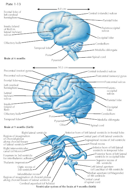
By sixth month of gestation, the cerebral hemispheres acquire several features that prefigure the division of the cortical surface into specific functional regions. Accordingly, one recognizes four distinct domains, or lobes, that define cortical territories. The frontal lobe is most anterior; it eventually includes cortical areas devoted to motor control, language production (left hemisphere only in most individuals), and executive function—the capacity, moment by moment, to integrate perceptions of external stimuli with internal representations of motivations, goals, and memories to plan appropriate complex behavioral responses. Midway along the anterior-posterior axis, the central (also known as the rolandic) sulcus divides the frontal and more posterior parietal lobe, which mediates somatosensation and attention. This anatomic landmark is one of the earliest local furrowings that defines the sulci (grooves) and gyri (bulges) that reflect the elaborate folding of the mature cerebral cortex.
The central sulcus defines two
essential functional regions. On the anterior bank
is the precentral gyrus, the location of the primary motor cortex. Neurons
of the primary motor cortex send axons directly to brainstem and spinal cord
motor neurons that innervate muscles or to interneurons adjacent to these motor
neurons. On the posterior bank is the postcentral gyrus, the location of
the primary somatosensory cortex. The primary somatosensory cortex
receives topographically mapped inputs from brainstem and spinal cord sensory
relay nuclei that represent somatosensory information from the entire body
surface. The remainder of the parietal lobe is devoted to sensory integration
and attention. Posterior and ventral, marked by the parieto-occipital
sulcus, is the occipital lobe, devoted exclusively to representation
and processing of vision. Finally, the anterior medial extension of the
hemisphere defines the temporal lobe, anterior to the lateral sulcus,
including cortical regions that integrate information about the identity of
visual stimuli, auditory information, and, in the left hemisphere only of most
individuals, the representation of “lexical” language (the brain’s “dictionary”
of words). The initial growth of the frontal, parietal, occipital, and temporal
lobes results in “operculation,” or covering of one region of cortical tissue
called the insula. The cortex of the insula becomes specialized for
visceral and homeostatic control and the representation of taste information.
The dramatic growth of the cerebral
hemispheres is accompanied by differentiation of the cerebellum and medulla. By
the end of the sixth month of gestation, the cerebellum expands with furrows
and ridges that eventually become the highly folded folia of the cerebellar
cortex (note: a cortex is the outer sheet of cells that invests any organ). The
pons is distinct, consisting of axons from the cortex that project to pontine
nuclei that then send axons to the cerebellar cortex. The medulla also becomes
furrowed and ridged, but for a different reason. The pyramid, a
prominent ridge on the anterior/ medial medulla is formed by growth of axons
from motor cortical neurons to the brainstem and spinal cord. By the end of
gestation, the pyramid is adjacent to a more lateral ridge, the olive. The
olive reflects accumulation of neurons into the olivary nucleus; olivary
neurons selectively innervate the extensive dendritic arbors of Purkinje cells.
Finally, further elaboration of the
ventricular system accompanies these morphogenetic transformations.
The ventricular system is best
depicted as a “cast” of space within the brain and spinal cord (neural tissue
is absent). The key changes reflect differential growth of brain regions that
correspond to each ventricular division. The lateral ventricles grow
disproportionately and acquire further anatomic definition. The anterior
horn extends into the frontal lobe, with the caudate nucleus of the basal
ganglia as its floor. The inferior horn extends into the temporal lobe;
on its anterior and medial surface is the hippocampus. Finally, the posterior
horn extends into the occipital lobe. The
remainder of the ventricular system is comprised of the same subdivisions
that emerge in the second trimester; however, their shape and size change
substantially. The third ventricle becomes a narrow midline space, further
indented by the thalamus on each side (the thalamic impression) as well
as a foramen surrounding the intrathalamic adhesion. In addition,
the third ventricle is indented by the optic chiasm at the optic recess, the
pineal gland at the pineal and suprapineal recess, and
the pituitary gland at the infundibular recess.


