POSITRON EMISSION TOMOGRAPHY
SCANNING
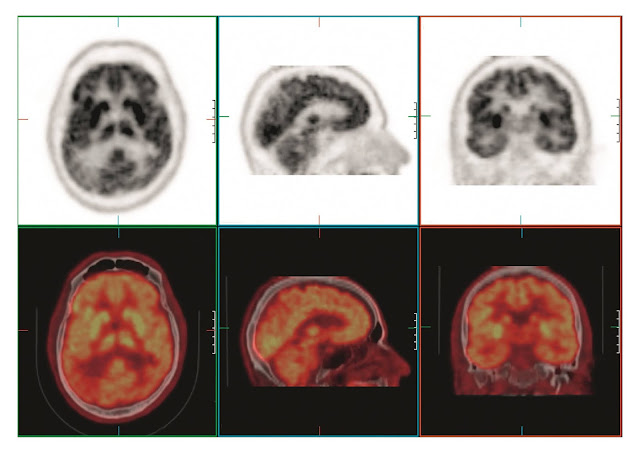
Positron emission tomography (PET) scanning is designed to assess the distribution of tracers labeled with positron-emitting nuclides, such as carbon-11 (11C), nitrogen-13 (13N), oxygen-15 (15O), and fluorine-18 (18F). Fluorodeoxyglucose (FDG), a glucose analogue labeled with 18F, can cross the blood-brain barrier. The metabolic products of FDG become immobile and trapped where the molecule is first used, thereby permitting FDG to be used to map glucose uptake in the brain.
This is a valuable tool for investigating subtle
physiological processes related to neurological
diseases. The distribution of FDG can be localized and reconstructed using
standard tomographic techniques that show the tracer distribution through- out
the body or brain. In this example of axial, sagittal, and coronal views, the
transmission measurement and correction was performed immediately following PET
acquisition using a 16-slice CT unit. The PET and CT images were automatically
fused by anatomical coregistration software (shown as colored images).


