MUSCULATURE OF STOMACH
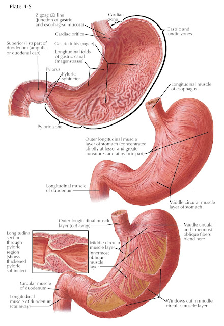
The musculature of the gastric wall consists solely of smooth muscle fibers, which, in contrast to the dual-layered arrangement elsewhere in the digestive tract, exist in three distinct layers. Only the middle circular layer covers the wall completely; the other two layers, the superficial longitudinal and deepest oblique layers, are present as incomplete coats. The longitudinal and circular layers are interconnected by continuous fibers, as are the circular layer and the deepest oblique muscular coats.
The longitudinal layer of the stomach is continuous with the longitudinal muscle layer of the esophagus, which divides at the cardia into two stripes. The stronger of these muscle bands follows the lesser curvature across the superior border of the stomach. The other set of fibers is somewhat broader but thinner as it migrates along the fundus, across the greater curvature toward the pylorus. Thus, the middle areas of the anterior and posterior surfaces of the stomach remain mostly free of longitudinal muscle fibers. The marginal muscle fibers of the upper longitudinal muscle stripe radiate obliquely toward the anterior and posterior surfaces of the fundus and corpus to unite with fibers of the circular layer. In the pyloric area, the two bands of longitudinal muscle fibers converge again to form a uniform layer, which, to a great extent is continuous with the external, longitudinal muscular layer of the duodenum. It is the increased thickness of the longitudinal muscle layer in the anterior and posterior parts of the pylorus that is responsible for the so-called pyloric ligaments (anterior and posterior pyloric ligaments, respectively).
The circular layer of smooth muscle is not only
the most continuous but the strongest of the stomach’s three layers. It
also begins at the cardia as the continuation of the superficial fibers of the
circular esophageal muscle. The circular layer becomes markedly more pronounced
as it approaches the pylorus, forming the pyloric sphincter.
The innermost layer is formed by oblique fibers of
the smooth muscle layer; it is most strongly developed in the region of the
fundus and becomes progressively weaker as it approaches the pylorus. In the
cardiac region, its fibers connect with the deeper circular layers of the
esophageal muscle. No oblique fibers exist in the vicinity of the lesser
curvature, but the fibers closest to it arise from a point to the left of the
cardia and run parallel to the lesser curvature. There are longitudinal furrows
in the lesser curvature caused by the absence of this innermost oblique layer.
The oblique fiber bundles, following these first more or less longitudinal
fibers, bend farther and farther to the left and, finally, become practically
circular in the region of the fundus, where their continuity with the fibers of
the circular layer is clearly evident. Because the oblique fibers of the
anterior and posterior walls merge into one another in the region of the
fundus, the oblique layer as a whole is made of U-shaped loops. The oblique
fibers never reach the greater curvature in the region of the corpus but fan
out and gradually disappear in the walls of the stomach. To the left of the
esophagus the oblique fibers
form sling fibers that project from the anterior wall of the stomach to
the posterior wall, making a tight bend around the cardial notch. On the
opposite side of the cardiac region, the circular layer has substantial clasp
fibers that pinch and narrow the cardiac region. Acting together, the
sling and clasp fibers help prevent gastric reflux and form a functional, but
not anatomic, lower esophageal sphincter.
MUSCULATURE OF PYLORIC SPHINCTER
The middle circular layer thickens considerably at the
pylorus, forming a muscular ring that acts as a true anatomic sphincter. The
pyloric sphincter is not continuous with the circular musculature of the
duodenum but is separated from it by a thin, fibrous septum of connective
tissue. A few fibers of the longitudinal muscle layer, the greater mass of
which, as mentioned above, is continuous with the corresponding layer of the
duodenum, contribute also to the muscle mass of the pyloric sphincter; they may
even find their way into the network of the sphincter bundles and penetrate as far as the
submucosa.


