MATURE BRAIN
VENTRICLES
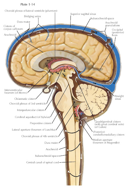
Few anatomic features of the mature brain reflect brain development more directly than the brain ventricles. This continuous system of fluid-filled chambers is the very same space that was defined by the closure of the neural tube. Subsequent morphogenesis modifies this space; nevertheless, its relationship to the original lumen of the neural tube is clear. CSF, which is pro- duced by the choroid plexus found in the lateral, third, and fourth ventricles, circulates throughout this space in the adult (as well as the embryonic) brain. The ventricular space also has a series of continuities with the subarachnoid space so that CSF is also bathing the external as well as deep (or ventricular) surface of the brain.
In the adult brain, the two mature
cerebral hemispheres surround the lateral ventricles. These two
ventricles, the largest of the ventricular chambers, have three extensions into
distinct regions of the cerebral hemispheres. The anterior horns extend
into the frontal lobes, the inferior horns into the temporal lobe
(including adjacent to the hippocampus) and the posterior horns into the
occipital lobes. The atrium is the junction of the anterior, posterior,
and temporal horns. The relationship between the anterior horns of the lateral
ventricles, the corpus callosum posteriorly, the caudate nucleus
anterolaterally, and the third ventricle and thalamus anteromedially is shown
in the lower panel. The lateral ventricles remain continuous with the third
ventricle via the intraventricular foramen of Monro (the white arrow at
left in the lower panel shows the continuity between lateral and third
ventricles provided by the foramen of Monro: there are two). The third
ventricle extends the anterior to posterior length of the diencephalon. Its
proximity to the optic chiasm and pituitary gland anteriorly results in local
“indentations” known as the optic and infundibular recesses.
Similarly, the relationship of the third ventricle to the pineal gland defines
the pineal and suprapineal recesses in the posterior aspect of
the third ventricle.
The third ventricle is continuous
with the cerebral aqueduct, which travels through the mature
mesencephalon. The cerebral aqueduct connects to the fourth ventricle, which
is adjacent to the cerebellum and pons, and extends
into the upper medulla. The fourth ventricle has a significant bilateral
extension, the lateral recess that opens into the inferior cerebellar
peduncle. The fourth ventricle also has several specialized continuities with
the subarachnoid space to facilitate the circulation and drainage of cerebrospinal
fluid, which maintains the integrity of cells at the ventricular zone and
also contributes to the stability of the ionic milieu in the brain tissue
generally. Thus the two lateral apertures (also known as the foramen of Luschka) are continuous
with the subarachnoid space at the lateral aspect of the pontocerebellar
junction (near the inferior cerebellar peduncle) and the median aperture,
located at the midline where the two lateral recesses originate, is continuous
with the cerebellomedullary cistern (also referred to as the cisterna
magna). Indeed, there is a distributed system of cisterns throughout the subarachnoid space that provide reservoirs of CSF.


