CRANIOSYNOSTOSIS
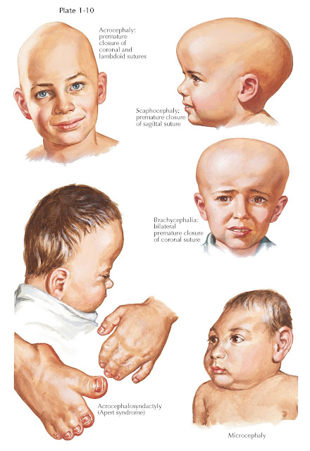
The growth of the brain is matched by the flexible growth of the cranial bones, which must establish a mechanism to expand the skull vault coincident with increased brain volume. The cranial bones, mostly generated by neural crest–derived chondrogenic and osteogenic precursors, are arranged as “plates” with elastic joints between each plate referred to as cranial sutures. Craniosynostosis implies a premature closure of one or more cranial sutures (see Plate 1-10). Early fusion of bone plates results in a progressively dysmorphic cranial shape. True craniosynostosis occurs in one of every 2,000 infants, predominates in males, and manifests in nonsyndromic and syndromic forms. Normally, the metopic, or frontal, suture closes before birth; the posterior fontanelle, at the union of the lambdoid and sagittal sutures, by 3 months; and the anterior fontanelle at the junction of the coronal, sagittal, and metopic sutures, by 18 months. After a suture is fused, growth occurs parallel to that suture; that is, growth is inhibited at 90 degrees to the suture. The fusion itself is felt as a ridge. Cranial sutures cannot be separated by increased intracranial pressure after 12 years of age.
Nonsyndromic craniosynostosis occurs
much more frequently than syndromic. The most common premature closure occurs
in the sagittal suture, which leads to scaphocephaly, dolichocephaly,
or elongated head. The next most common premature closure is found in the coronal
suture, which may be either unilateral or bilateral. If unilateral, it
causes a unilateral ridge, with a pulling up of the orbit, flattening of the
frontal area, and prominence near the zygoma on the affected side, which
produces a quizzical expression. If premature coronal closure is bilateral, brachycephalia,
manifested by an abnormally broad skull, is the result. Metopic craniosynostosis causes trigonocephaly, with a pointed frontal bone,
hypotelorism, and prominent temporal hollowing. True lambdoid synostosis, which
can also be unilateral or bilateral, is exceedingly rare, with an incidence
less than 1:100,000. Turricephaly, a towering cranial vault due to
multiple suture closure, is quite rare and disfiguring. Some infants will have
prominent ridges along sutures without the other typical cranial findings, and
these ridges will spontaneously resolve with time.
Syndromic craniosynostosis usually
is autosomal dominant. Crouzon disease, with closure of multiple sutures and
the associated facial anomalies of hypertelorism, proptosis, and choanal
atresia, is known as craniofacial dysostosis. Intelligence is normal,
but premature suture closure can cause elevated intracranial pressure. In acrocephalosyndactyly,
or Apert syndrome, the head is elongated, the result of premature closure of
all sutures; the orbits are shallow, causing exophthalmos; and either
syndactyly or polydactyly is present. Saethre-Chotzen, Pfeiffer, and Carpenter
have also identified syndromes of acrocephalosyndactyly that include various
combinations of synostosis, syndactyly, and other anomalies. Syndromic
craniosynostosis can be associated with hydrocephalus.
Conditions that can be confused with
craniosynostosis include microcephaly and deformational plagiocephaly.
Microcephaly from lack of brain growth is not typically accompanied by a
disfigured cranial shape. Deformational plagiocephaly is very common and
currently occurs in approximately 1 in 10 infants. The baby tends to lie on one
area of the skull, which causes flattening in the affected
area, anterior displacement of the unilateral ear, and ipsilateral frontal and
contralateral parietal bossing, with an overall parallelogram shape. Some
infants have associated torticollis. Most infants will have spontaneous
improvement with exercises; very severe cases made need treatment with a
cranial orthosis.
The diagnosis of craniosynostosis is
made by clinical examination in most cases. Appropriate radiographic examinations are typically only needed as a
roadmap for surgical repair. Treatment for true craniosynostosis is surgical,
with either endoscopic or open techniques. Early referral optimizes the
opportunity to use minimally invasive techniques. Treatment of syndromic and
multiple suture craniosynostosis typically require multiple procedures by an
experienced craniofacial team during early
childhood.


