ANATOMY OF THE THORACOLUMBAR
AND SACRAL SPINE
THORACIC
VERTEBRAE AND LIGAMENTS
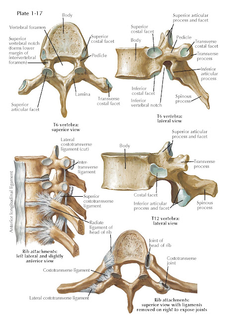
The 12 thoracic vertebrae (T1 to T12) are intermediate in size between the smaller cervical and the larger lumbar vertebrae. The heart-shaped vertebral bodies are slightly taller posteriorly than anteriorly, producing a slight wedge shape (see Plate 1-17). Vertebrae are easily recognized by their costal facets on both sides of the bodies and on all the transverse processes except those of T11 and T12. The costal facets articulate with the facets on the heads and tubercles of the corresponding ribs. The spinal canal is smaller and more rounded than in the cervical spine and corresponds to the more circular shape of the spinal cord in the thoracic region. The spinal canal is formed by the posterior surfaces of the vertebral bodies and by the pedicles and laminae forming the vertebral arches. The stout pedicles are directed posteriorly; they have shallow superior and much deeper inferior vertebral notches. The laminae are short and relatively thick and partially overlap each other from above downward.
The typical thoracic superior articular processes project upward from
the junction of the pedicles and the laminae, and their facets slant
posteriorly and slightly upward and outward. The inferior articular processes
project downward from the anterior parts of the laminae, and their facets face
forward and slightly downward and inward.
 |
THORACIC VERTEBRAE AND LIGAMENTS
Most of the thoracic spinous processes are long and inclined inferiorly and posteriorly. Those of the upper and lower thoracic vertebrae are more horizontal. The transverse processes are also relatively long and extend posterolaterally to form the junctions of the pedicles and laminae. Except for the lowest two (or occasionally three) thoracic vertebrae, the transverse processes have small oval facets near their tips that articulate with similar facets on their corresponding rib tubercles.
Adjacent vertebral bodies are connected by the intervertebral discs as
well as by the anterior and posterior longitudinal ligaments; the transverse
processes are connected by the intertransverse ligaments; the laminae are
connected by the ligamentum flavum; and the spinous processes are connected by
the supraspinous and interspinous ligaments. The facet joints, which form from
adjacent superior and inferior articular processes, are synovial joints and are
covered by a fibrous articular capsule.
Costovertebral Joints
The ribs are connected to the vertebral bodies and the transverse
processes by various ligaments. The costocentral joints, between the bodies and
the rib heads, have articular capsules. The second to tenth costal heads, each
of which articulates with two vertebrae, are also connected
to the corresponding vertebral discs by interarticular ligaments. Radiate
(stellate) ligaments unite the anterior aspects of the rib heads with the sides
of the vertebral bodies above and below the discs. The rib head articulates
with both the upper border of its own vertebra as well as the lower border of
the vertebra above (i.e., the ninth rib articulates with the vertebral bodies
of both T8 and T9). The costotransverse joints between the facets on the
transverse processes and on the tubercles of the ribs are also surrounded by
articular capsules. They are reinforced by a (middle) costotransverse ligament
between the rib neck and transverse process of the vertebra above and a lateral
costotransverse ligament interconnecting the end of a transverse process with
the nonarticular part of the related costal tubercle.
LUMBAR VERTEBRAE AND INTERVERTEBRAL DISCS
LUMBAR VERTEBRAE AND INTERVERTEBRAL DISCS
The five lumbar vertebrae (L1 to L5) are the largest separate vertebrae
and are distinguished by the absence of costal facets (see Plate 1-18). The
vertebral bodies are wider from side to side than from front to back, with
upper and lower surfaces that are kidney shaped and almost parallel, except in
the case of the fifth vertebral body, which is slightly wedge shaped. The
vertebral foramina have a “teardrop” shape. The pedicles are short and strong,
arising from the upper and posterolateral aspects of the bodies. The laminae
are short, broad plates that meet in the middle to form nearly horizontal
spinous processes that are perpendicular to the lamina.
The intervals between adjacent laminae and spinous processes (interlaminar
spaces) are wider than in the cervical and thoracic spine.
The articular processes (which form the facet joints) project superiorly
and inferiorly from the area between the pedicles and laminae. The facet joints
are oriented relatively vertically, which allows for some flexion and extension
but little rotation. The transverse processes of L1 to L3 are long and slender,
whereas those of L4 and L5 are more pyramidal. Near the base of each
transverse process are small accessory processes; other small, rounded
mammillary processes protrude from the posterior margins of the superior
articular processes.
The fifth lumbar vertebra is atypical. It is the largest, with its body
deeper anteriorly; its inferior articular facets face almost forward and are
set more widely apart, and the roots of its stumpy transverse processes are
continuous with the entire lateral surfaces of the pedicles.
Interposed between the adjacent vertebral bodies are intervertebral
discs. These are immensely strong fibrocartilaginous structures that provide
powerful bonds between vertebrae and act to buffer axial loads. The discs
consist of outer concentric layers of fibrous tissue, the anulus fibrosus. The
fibers in adjacent layers of the anulus are arranged obliquely but in opposite
directions to resist torsion. The anulus fibrosus contains a central, springy
and pulpy zone called the nucleus pulposus. The vascular and nerve supply to
the discs is minimal. If the fibers of the annulus fail as a result of injury
or disease, the enclosed nucleus pulposus may extrude posteriorly through the
rent in the annulus and press on adjacent neural structures.
In the normal spine, the discs account for almost 25% of the height of the
vertebral column; they are thinnest in the upper thoracic region and thickest
in the lumbar region. In the vertical section, the lumbar discs are moderately
wedge shaped, with the thicker edge anteriorly. The convexity (lordosis) of the
normal lumbar spine is due more to the shape of the discs than to the shape of
the vertebral bodies. With aging, the nucleus pulposus undergoes changes: its
water content decreases, its mucoid matrix is gradually replaced by
fibrocartilage, and it ultimately comes to resemble the anulus fibrosus.
Although the resultant loss of height in each disc is small, it may amount to
an overall decrease of 2 to 3 cm in the length of the vertebral column.
SACRAL SPINE AND PELVIS
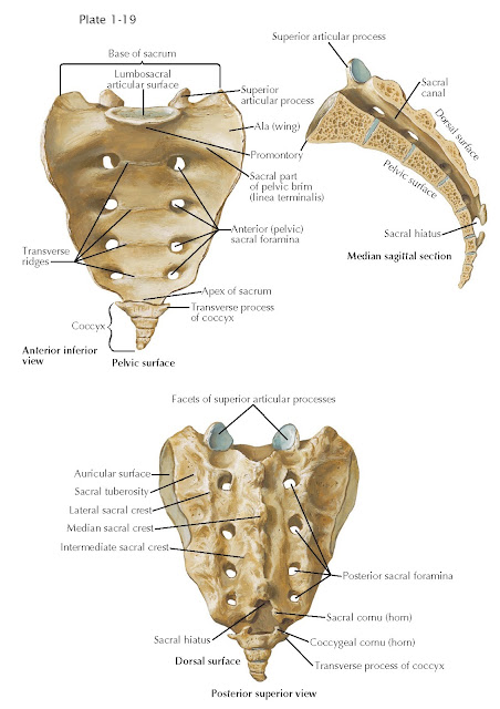
The principal function of the pelvis is to transmit body weight to the
limbs and absorb the muscular stresses of an upright posture (see Plate 1-19). The center
of gravity of the body passes just anterior to the sacral promontory.
The sacrum, composed of five fused vertebrae, broadens laterally into
the sacral ala. The sacral nerve roots emerge through the anterior and
posterior sacral foramina just medial to the sacral ala. The anterior surface
of the sacrum is smooth. Dorsally, the surface is highly irregular to
facilitate ligamentous attachments. The lateral articular surfaces of the
sacrum articulate with the pelvis, forming the sacroiliac joint. The joint
surfaces contain complementary elevations and depressions to diminish motion.
Additionally, the anterior and posterior sacroiliac
joint capsules have overlying sacroiliac ligaments, which are among the
strongest in the body.
LUMBOSACRAL LIGAMENTS
The anterior longitudinal ligament is a straplike band that increases in
width, moving caudally down the spine. It extends from the anterior tubercle of
the atlas to the sacrum. It is firmly attached to the anterior margins of the
vertebral bodies and discs (see Plate 1-20). The posterior longitudinal ligament is broader in
the upper spine and becomes narrower as it traverses caudally. It lies within
the vertebral canal immediately behind the vertebral bodies. Its upper end is
continuous with the tectorial membrane, and it extends from the axis to the
sacrum. The edges of the ligament are serrated, particularly in the lower
thoracic and lumbar regions, because it spreads out between its attachments to
the vertebral bodies to blend with the annular fibers of the discs. The ligament
is separated from the posterior vertebral body surfaces by basivertebral veins
that join the anterior internal venous plexus. The posterior longitudinal
ligament is stronger in the midline and weaker laterally, which helps to
explain the propensity for lumbar disc herniations to occur in a
posterolateral, rather than midline, location.
 |
| LUMBOSACRAL LIGAMENTS |
The ligamentum flavum is largely composed of yellow elastic tissue and
joins adjacent laminae. It extends from the anteroinferior surface of the
lamina above to the posterosuperior aspect of the lamina below, and from the
midline to the facet joint capsules laterally. There is a midline raphe
separating the right and left ligaments. The ligaments increase in thickness
from the cervical to the lumbar spine.
The supraspinous ligaments connect the tips of the spinous processes
from the seventh cervical vertebra to the sacrum. They are continuous with the
nuchal ligament in the cervical spine and the interspinous
ligaments immediately anterior, and they increase in thickness caudally. The
interspinous ligaments are thin, membranous structures between the roots and
apices of the spinous processes and are best developed in the lumbar region.
The interosseous sacroiliac ligaments are formed by short, thick bundles
of fibers connecting the sacral and iliac tuberosities. The dorsal sacroiliac
ligaments fill the deep depression between the sacrum and the iliac bones
dorsally. The sacrotuberous ligament is long, flat, and triangular. It arises
from the posterior superior and posterior inferior iliac spines and from the
back and side of the sacrum. The fibers converge on the ischial tuberosity. The
sacrospinous ligament arises from the lateral aspect of the lower sacrum and
coccyx and attaches to the ischial spine. This ligament converts the greater
sciatic notch into the greater sciatic foramen and with the
sacrotuberous ligament forms the lesser sciatic foramen. The lumbosacral joint
unites L5 and the sacrum. These vertebra are united by
the same ligamentous structures found throughout the lumbar spine with the
addition of the strong iliolumbar ligament traversing laterally from the
transverse process of L5 to the posterior part of the iliac crest. The
iliolumbar ligament resists the tendency of the lumbar vertebra to slip down the slope of
the sacral promontory.


