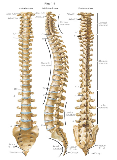
VERTEBRAL
COLUMN
The vertebral column is built from individual units of alternating bony vertebrae and fibrocartilaginous discs. These units are intimately connected by strong ligaments and supported by paraspinal muscles with tendinous attachments to the spine. The individual bony elements and ligaments are described in Plates 1-9 to 1-18.
There are 33 vertebrae
(7 cervical, 12 thoracic, 5 lumbar, 5 sacral, and 4 coccygeal), although the
sacral and coccygeal vertebrae are usually fused to form the sacrum and coccyx.
All vertebrae conform to a basic plan, but morphologic variations occur in the
different regions. A typical vertebra is made up of an anterior, more-or-less
cylindrical body and a posterior arch composed of two pedicles
and two laminae, the latter united posteriorly in the midline to
form a spinous process. These processes vary in shape, size, and
direction in the various regions of the spine. On each side, the arch also
supports a transverse process and superior and inferior articular
processes; the latter form synovial joints that are the posterior sites of
contact (left and right) for adjacent vertebral segments. The disc is the
anterior site of attachment. The spinous and transverse processes provide
levers for the many muscles attached to them. The increasing size of the
vertebral bodies from above downward is related to the increasing weights and
stresses borne by successive segments, and the sacral vertebrae are fused to
form a solid wedge-shaped base—the keystone in a bridge whose arches curve down
toward the hip joints. The intervertebral discs act as elastic buffers
to absorb the many mechanical shocks sustained by the vertebral column.
Only limited movements
are possible between adjacent vertebrae, but the sum of these movements confers
a considerable range of mobility on the vertebral column as a whole. Flexion,
extension, lateral bending, rotation, and translation are all possible, and
these actions are freer in the cervical and lumbar regions than in the thoracic
region. Such differences exist because the discs are thicker in the cervical
and lumbar areas and they lack the splinting effect produced by the thoracic
rib cage and sternum. Additionally, the cervical and lumbar spinous processes
are shorter and less closely apposed and the articular processes are shaped and
arranged differently.
At birth, the
vertebral column presents a general dorsal convexity, but, later, the cervical
and lumbar regions become curved in the opposite directions when the infant
reaches the stages of holding up the head (3 to 4 months) and sitting upright
(6 to 9 months). The dorsal convexities are primary curves associated
with the fetal uterine position, whereas the cervical and lumbar ventral secondary
curves are compensatory to permit the assumption of the upright position.
There may be additional slight lateral deviations resulting from unequal
muscular traction in right-handed and left-handed persons.
The evolution of the
human from a quadrupedal to a bipedal posture has been mainly attributed to the
tilting of the sacrum between the hip bones, by an increase in
lumbosacral angulation, and by minor adjustments of the anterior and posterior
depths of various vertebrae and discs. An erect posture greatly increases the
load on the lower spinal joints; however, as good as these ancestral
adaptations were, some static and dynamic imperfections remain and predispose
to the effects of gradual strain.
The length of the
vertebral column averages 72 cm in the adult male and 7 to 10 cm less in the
female. The vertebral canal extends through the
entire length of the column and provides an excellent protection for the spinal
cord, the exiting nerve roots, and the cauda equina. Vessels and nerve roots
pass through intervertebral foramina between the superior and inferior
borders of the pedicles of adjacent vertebrae, bound anteriorly by the
corresponding vertebral body and intervertebral discs and posteriorly by the
joints between the articular processes of
adjoining vertebrae.


