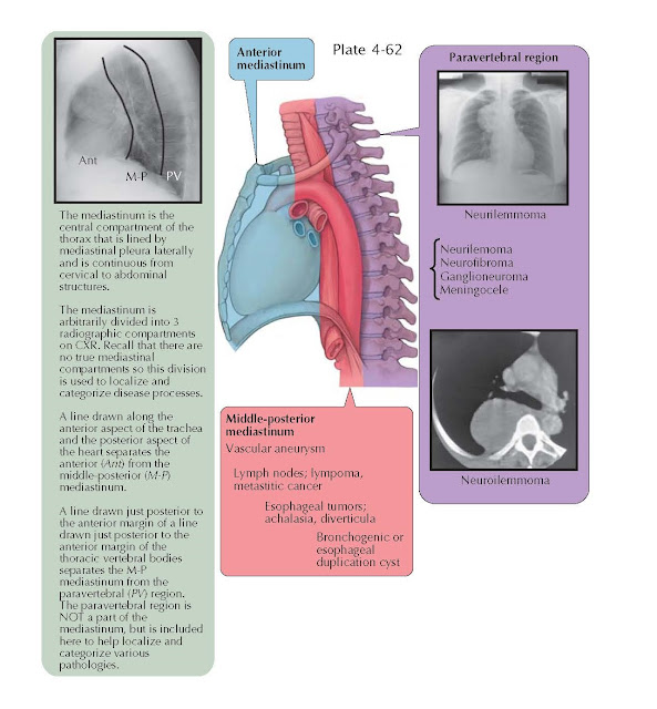
MIDDLE-POSTERIOR AND PARAVERTEBRAL
MEDIASTINUM
The middle-posterior mediastinum is the compartment located between the
anterior and the paravertebral compartments. The paravertebral compartment is
located posterior to an imaginary line drawn just posterior to the anterior
margins of the thoracic vertebral bodies. Approximately 10% to 15% of
mediastinal tumors occur in the middle-posterior mediastinum. The most common
abnormalities are caused by lymph node enlargement from lymphoma, granulomatous
disease caused by tuberculosis, fungal infection, or non-infectious conditions
of sarcoidosis or silicosis. Metastatic lymphadenopathy may be caused by
cancers of the lung, kidney, breast, or gastrointestinal tract. Rare causes of
lymphadenopathy are Castleman disease and amyloidosis. Symptoms may be absent
or related to the underlying systemic disease process, such as fever and night
sweats caused by lymphoma or an infectious process. Some patients may complain
of dysphagia caused by compression of the esophagus or vague chest discomfort.
Congenital foregut cysts are a common cause of middle
mediastinal lesions. These include bronchogenic cysts, esophageal duplication
cysts, and (uncommonly) neurenteric cysts. Bronchogenic cysts are most common
and are thought to be caused by an abnormal budding of the foregut during
development. Most are located paratracheally or subcarinally. These cysts are
lined by respiratory epithelium (pseudostratified, columnar, ciliated). Enteric
cysts (esophageal duplication and neurenteric) arise from the dorsal foregut
and are usually located in the middle-posterior mediastinum. Enteric cysts are
lined by squamous or enteric epithelium. The cyst walls have smooth muscle
layers with a myenteric plexus. Esophageal duplication cysts usually adhere to
the esophagus. Neurenteric cysts may be associated with the esophagus or
cervical or upper thoracic vertebral abnormalities with an attachment or
extension into the spine. Most enteric cysts are diagnosed during childhood.
Radiographically, these cysts are well-circumscribed, spherical masses. On
computed tomography (CT) scan, they are unilocular, homogeneous, and
nonenhancing.
Asymptomatic cysts may be observed, but their chance
or rate of enlargement is uncertain. Large cysts may compress the airways and
lead to pneumonia or dysphagia with esophageal compression. The treatment of
choice for patients with symptomatic cysts is surgical resection. Unresected
cysts rarely transform into malignant lesions.
Esophageal disorders such as achalasia, benign tumors,
diverticula, and carcinoma are common causes of middle-posterior mediastinal
lesions. Hiatal hernia is very common and presents as a mass in the inferior
middle-posterior mediastinum, usually seen as a retro- cardiac mass on routine
chest radiography. Barium swallow studies and endoscopy are generally
diagnostic. Vascular lesions may present as a mass in the middle-posterior
mediastinum and should always be considered before attempting biopsy. Thoracic
aortic aneurysm is the most common of these, but pulmonary artery aneurysm and
mediastinal hemangiomas are occasionally encountered. Contrast-enhanced CT
chest examinations are generally diagnostic for a vascular aneurysm and likely
demonstrate the vascular nature of hemangiomas.
Tumors of the paravertebral compartment are generally
caused by neurogenic neoplasms. Neurogenic tumors account for 20% of adult and
40% of pediatric mediastinal tumors. The large majority of these tumors in
adults are benign, but 50% of the neurogenic tumors in children are malignant.
Schwannomas (also called neurilemmomas) and neurofibromas are the most
common neurogenic neoplasms. More than 90% are benign, and a small percentage
are multiple. They are slow growing and arise from a spinal nerve root. Neurofibromas
often occur in individuals with von Recklinghausen disease (neurofibromatosis).
They may have multiple tumors, and malignant transformation is more common with
this disease. These tumors are most commonly asymptomatic and detected
accidentally. Occasionally, they may result in pain that leads to discovery of
the tumor. The malignant form of these neurogenic tumors is classified as a
malignant peripheral nerve sheath neoplasm. Radiographically, schwannomas and
neurofibromas are well-marginated, spherical, or lobular paravertebral masses.
They are usually small and span one to two vertebrae but can grow to a large
size. They may cause erosion of the rib or vertebral body, and 10% grow through and enlarge the
neuroforamina and expand on either end to give a “dumbbell” shape. For this
reason, magnetic resonance imaging of the spine is indicated before surgical
resection is attempted. Surgery is the treatment of choice.
Ganglioneuromas are benign neoplasms of the
sympathetic ganglia that typically occur in older children or young adults.
They may be asymptomatic or symptomatic because of local tumor effects. They
are well-demarcated, oblong paravertebral masses that usually span three to five
vertebrae. Ganglioneuroblastomas and neuroblastomas are malignant sympathetic
ganglia neoplasms that occur in young children.
Pheochromocytomas or paraganglionomas are rare
neoplasms of paraganglionic tissue and rarely occur in the mediastinum, usually
adjacent to the aorta or pulmonary arteries, but they may present in the
posterior mediastinum. A lateral thoracic meningocele is uncommon and may be
multiple. Radiographic studies demonstrate the typical cystic structures in the
paravertebral foramina location. Asymptomatic lesions do not require treatment.


