JOINTS OF THE HAND
The carpometacarpal joint of the
thumb is the independent joint between the trapezium and the base of the first
metacarpal (see Plate 4-3). The articular surfaces are reciprocally
concavoconvex, and a loose but strong articular capsule joins the bones. The
biaxial nature of this joint provides for flexion and extension and abduction
and adduction, and the looseness of its capsule allows opposition of the thumb
that involves a small amount of rotary movement (circumduction).
The carpometacarpal joints of the
four fingers participate with the intercarpal and intermetacarpal joints in a
common synovial cavity. Dorsal and palmar carpometacarpal ligaments run from
the carpals of the second row to the various metacarpals. Short interosseous
ligaments are usually present between contiguous angles of the capitate and the
hamate and the third and fourth metacarpals.
INTERMETACARPAL JOINTS
These joints occur between the
adjacent sides of the bases of the four metacarpals of the fingers. Here, also,
there are dorsal and palmar ligaments, and interosseous ligaments close off the
common synovial cavity by connecting the bones just distal to their articular
facets. Only slight gliding movements occur between the metacarpals and between
them and the carpals to which they are related. However, the articulation
between the hamate and the fifth metacarpal allows that bone to flex
appreciably during a tight grasp and also to rotate slightly
under the traction of the opponens digiti minimi muscle.
The deep transverse metacarpal
ligaments are short and connect the palmar surfaces of the heads of the
second, third, fourth, and fifth metacarpals. They are continuous with the
palmar interosseous fascia and blend with the palmar ligaments of the
metacarpophalangeal joints and the fibrous sheaths of the digits. They limit
the spread of the metacarpals, and the tendons of the interosseous
and lumbrical muscles pass on either side of them.
METACARPOPHALANGEAL JOINTS
These joints are condyloid, and
both the rounded head of the metacarpal and the oval concavity of the proximal
end of the phalanx have unequal curvatures along their transverse and vertical
axes. An articular capsule and collateral and palmar ligaments unite the
bones. The articular capsule is rather loose. Dorsally, it is reinforced by the
expansion of the digital extensor tendon. The palmar ligament is a
dense, fibrocartilaginous plate, which, by means of its firm attachment to the
proximal palmar edge of the phalanx, extends and deepens the phalangeal
articular surface. It is loosely attached to the neck of the metacarpal; in
flexion, it passes under the head of the metacarpal and serves as part of the
articular contact of the bones. At its sides, the palmar ligament is continuous
with the deep transverse metacarpal ligaments and the collateral ligaments. The
collateral ligaments are strong, cordlike bands attached proximally to
the tubercle and adjacent pit of the head of the metacarpals and distally to
the palmar surface of the side of the phalanx. Their fibers spread fanlike to
attach to the palmar ligaments. Movements of flexion and extension, abduction
and adduction, and circum- duction are permitted at these joints. With
extension is associated abduction, as in fanning the fingers; with flexion is
associated adduction, as in making a fist. The metacarpophalangeal joint of the
thumb is limited in abduction and adduction; its special freedom of motion derives
from its carpometacarpal joint.
INTERPHALANGEAL JOINTS
Structurally similar to the
metacarpophalangeal series, the interphalangeal joints have the same loose
capsule, palmar and collateral ligaments, and dorsal reinforcement from the
extensor expansion. However, owing to the pulley-like form of their articular
surfaces, action here is limited to flexion and extension.
Flexion is freer than extension and may reach 115 degrees at the proximal interphalangeal
joint. Arteries and nerves serving these joints are twigs of adjacent proper
digital branches.
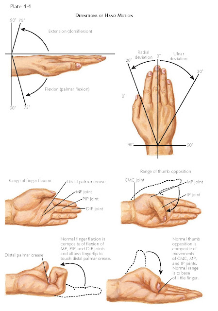
MOVEMENT OF THE HAND
The unique bony and articular
anatomy of the hand allow for a myriad of movements, and the cumulative movement
of each joint in series increases the total active motion (TAM). By convention,
movement toward the palm is described as palmar or volar or anterior and
movement toward the back of the hand is described as dorsal or posterior.
Movement of the hand toward the thumb side of the arm is described as radial or
lateral and toward the small finger as ulnar or medial.


