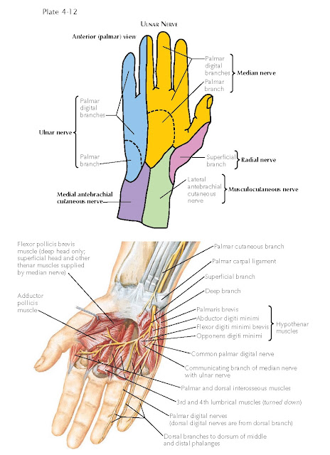
INNERVATION OF THE HAND
Nerve branches from the ulnar, median, and radial nerve supply motor,
sensory, and autonomic vasomotor function in the hand.
ULNAR NERVE
The ulnar nerve (C[7], 8; T1) is the main continuation
of the medial cord of the brachial plexus. In the forearm and hand, the ulnar
nerve gives off articular, muscular, palmar, dorsal, superficial and deep
terminal, and vascular branches. It divides into branches for the areas of skin
on the medial side of the back of the hand and fingers (see Plate 4-12). The ulnar nerve enters the hand to the radial side of the
pisiform between the palmar carpal ligament and the flexor retinaculum. Just
distal to the pisiform, the ulnar nerve divides into superficial and deep
branches.
The superficial terminal branch supplies the
palmaris brevis muscle, innervates the skin on the medial side of the palm, and
gives off two palmar digital nerves. The first is the proper palmar digital
nerve for the medial side of the small finger; the second, the common
palmar digital nerve, communicates with the adjoining common palmar digital
branch of the median nerve before dividing into the two proper palmar
digital nerves for the adjacent sides of the small and ring fingers.
Rarely, the ulnar nerve supplies two and one-half rather than one and one-half
digits, and the areas supplied by the median and radial nerves are reciprocally
reduced. The deep terminal branch of the ulnar nerve, with the deep
branch of the ulnar artery, sinks between the origins of the abductor digiti
minimi and the flexor digiti minimi brevis muscles and perforates the origin of
the opponens digiti minimi muscle. It supplies these muscles and then curves
around the hamulus of the hamate into the central part of the palm of the hand
in conjunction with the deep palmar arterial arch. As it crosses the hand deep
to the flexor tendons to the digits, the nerve gives twigs to the ulnar two lumbrical
muscles and to all the interosseous muscles, both dorsal and palmar. It then
supplies the adductor pollicis muscle and gives articular twigs to the wrist
joint, and it may send a
terminal branch into the deep head of the flexor pollicis brevis muscle.
Variations in the nerve supplies of the palmar muscles
are as common as the variations in the cutaneous distribution; they are due to
the variety of interconnections between the ulnar and median nerves.
The dorsal branch of the ulnar nerve completes the
cutaneous supply of the dorsum of the hand and digits. It arises about 5 cm
above the wrist, passes dorsalward from beneath the flexor carpi ulnaris tendon, and then pierces the
forearm fascia. At the ulnar border of the wrist, the nerve divides into three
dorsal digital branches. There are usually two or three dorsal digital
nerves, one supplying the medial side of the small finger, the second
splitting into proper dorsal digital nerves to supply adjacent sides of
the small and ring fingers, and the third (when present) supplying contiguous sides of the ring and long fingers.
The first branch
courses along the ulnar side of the dorsum of the hand and supplies the ulnar
side of the small finger as far as the root of the nail. The second branch
divides at the cleft between the ring and small fingers and supplies their
adjacent sides. The third branch may divide similarly; it may supply the
adjacent sides of the long finger and ring finger, or it may simply anastomose
with the fourth dorsal digital branch of the superficial branch of the radial
nerve. The dorsal branches to the ring finger usually extend only as far as the
base of the second phalanx, with the more distal parts of the ring and small
finger supplied by palmar digital branches of the ulnar nerve.
The palmar branch of the ulnar nerve arises
about the middle of the forearm, descending under the ante brachial fascia in front of the ulnar artery. It
perforates the fascia just above the wrist and supplies the skin of the
hypothenar eminence and the medial part of the palm.
MEDIAN NERVE
The median nerve (C[5], 6, 7, 8; T1) is formed by the
union of medial and lateral roots arising from the corresponding
cords of the brachial plexus (see (Plate 1-18).
The palmar branch of the median nerve arises just above the wrist (see Plate 4-13). It perforates the palmar carpal ligament between the tendons of the
palmaris longus and flexor carpi radialis muscles and distributes to the skin
of the central depressed area of the palm and the medial part of the thenar
eminence.
The digital branches of the median nerve, the
proper palmar digital nerves, lie subcutaneously along the margins of each of
the digits distal to the webs of the fingers (see Plates 4-12 and 4-13). They
arise from common palmar digital nerves, which lie under the dense palmar
aponeurosis of the central palm. The first common palmar digital nerve gives
rise to the muscular branch to the short muscles of the thumb and then divides
into three proper palmar digital nerves. Just distal to the flexor
retinaculum, its motor, or recurrent, branch curves sharply into
the thenar eminence and supplies the abductor pollicis brevis, flexor pollicis
brevis (sometimes
only its superficial head), and opponens pollicis muscles. This branch
frequently arises from the median nerve together with its first common digital
branch. The first common digital branch then runs to the radial and ulnar sides
of the thumb, giving numerous branches to the pad and small, dorsally running
branches to the nail bed of the thumb. The third proper digital branch supplies
the radial side of the index finger. The second common palmar digital branch provides two proper palmar digital nerves, which reach
the adjacent sides of the index and long fingers. The third common palmar
digital nerve communicates with a digital branch of the ulnar nerve in the palm
and divides into two proper palmar digital nerves supplying adjacent sides of
the long finger and ring fingers.
Proper palmar digital nerves are large because of the
density of nerve endings in the fingers. They lie superficial to the corresponding
proper palmar digital arteries and veins. As each nerve passes toward its termination in the
pad of the finger, it gives off branches for the innervation of the skin of the
dorsum of the digits and the matrices of the fingernails. These dorsal branches
innervate the dorsal skin of the distal segment of the index finger, the two
terminal segments of the long finger, and the radial side of the ring finger.
The common and proper palmar digital nerves vary in their origins
and distributions, but the usual arrangement innervates the skin (including the
nail beds) over the distal and dorsal aspects of the lateral three and one-half
digits. Occasionally, they supply only two and one-half digits. The proper
palmar digital branches to the radial side of the index finger and to the
contiguous sides of the index and long fingers also carry motor fibers to
supply the first and second lumbrical muscles, respectively. Therefore, the
digital nerves are not concerned solely with cutaneous sensibility. They
contain an admixture of efferent and afferent somatic and autonomic fibers,
which transmit impulses to and from sensory endings, vessels, sweat glands, and
arrectores pilorum muscles and between fascial, tendinous, osseous, and
articular structures in their areas of distribution.
RADIAL NERVE
Dorsally the superficial branch pierces the deep
fascia and commonly subdivides into two branches, which usually split into four
or five dorsal digital nerves. The cutaneous area of supply is shown in Plate 4-14. The smaller lateral
branch supplies the skin of the radial side and eminence of the thumb and
communicates with the lateral antebrachial cutaneous nerve. The larger medial
branch divides into four dorsal digital nerves. The first dorsal digital
nerve supplies the ulnar side of the thumb; the second supplies the radial side
of the index finger; the third distributes to the adjoining sides of the index
and long fingers; and the fourth supplies the adjacent sides of the long and
ring fingers.
There is usually an anastomosis on the back of the
hand between the superficial branch of the radial nerve and the dorsal branch
of the ulnar nerve, and there is some variability in the apparent source of the last (more median) branch
of either nerve. In some such cases, the adjacent sides of the long and ring
fingers are in the territory of the ulnar nerve. Dorsal digital nerves fail to
reach the extremities of the digits. They reach to the base of the nail of the
thumb, to the distal interphalangeal joint of the index finger, and not quite
as far as the proximal interphalangeal joints of the long and ring fingers. The
distal areas of the dorsum of the digits not supplied by the radial nerve receive branches from the stout palmar
digital branches of the median nerve.
The dorsal digital nerves also supply filaments to the
adjacent vessels, joints, and bones. (Note that the dorsal digital nerves
extend only to the levels of the distal interphalangeal joints and that the
first dorsal digital nerve gives off a twig that curves around the radial side
of the thumb to supply the skin over the lateral part of the thenar eminence.)


