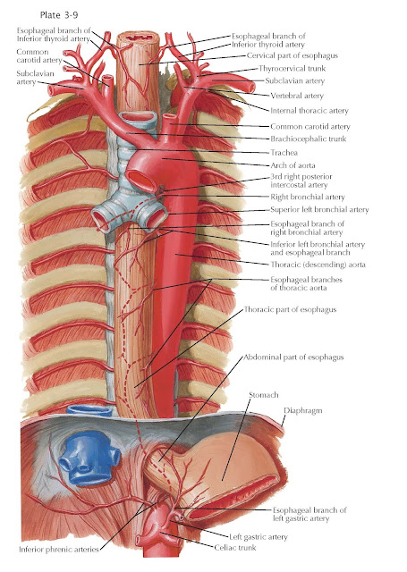
Blood Supply of Esophagus
The blood supply of the esophagus is extremely varied. The cervical
region of the esophagus receives blood from esophageal branches of the inferior
thyroid artery. The majority of esophageal branches arise from the terminal
branches of this artery; its ascending and descending portions frequently give
rise to one or more esophageal branches. The esophageal branches on the
anterior aspect of the esophagus give small branches to the nearby trachea.
Accessory arteries to the cervical esophagus may arise from the subclavian,
common carotid, vertebral, ascending pharyngeal, superficial cervical, and
thyrocervical arterial trunk.
The thoracic segment of the
esophagus is supplied by branches from the (1) bronchial arteries, (2) thoracic
aorta, and (3) right intercostal arteries. The bronchial arteries
give off esophageal branches at or below the tracheal bifurcation,
contributions from the left inferior bronchial artery being the most
common. Patterns of the bronchial arteries vary markedly. The standard text
book type (two left, one right) occurs only in about one half of persons.
Aberrant types are one right and one left (25%), two right and two left (15%),
one left and two right (8%), and, in some instances, three right or three left.
Near the bifurcation point of the trachea, the esophagus may receive additional
twigs from the abdominal aorta, aortic arch, and intercostal and internal
thoracic arteries. The aortic branches to the thoracic esophagus are not
segmentally arranged, nor are they four in number, as commonly taught, but are
only two unpaired vessels. The superior esophageal branch of the thoracic
aorta is short (3 to 4 cm) and usually arises at the level of T6 to T7. The
inferior esophageal branch of the thoracic aorta is longer (6 to 7 cm)
and arises at the T7 to T8 disk level. Both arteries pass posterior to the
esophagus and divide into ascending and descending branches that anastomose
longitudinally, with descending branches from the inferior thyroid artery as
well as bronchial arteries and with ascending branches from the left gastric
and left inferior phrenic arteries. Right intercostal arteries, mainly the
fifth, give rise to esophageal branches in about 20% of the population.
The abdominal esophagus receives
its blood supply primarily through branches that arise from the celiac trunk.
The left gastric artery is one of the three typical branches of this trunk and
is the major blood supply to the abdominal esophagus. An additional blood
supply comes from the short gastric arteries and from the recurrent
branch of the left inferior phrenic artery, given off by the latter after
it has passed posterior to the esophagus in its course to the diaphragm. The
left gastric artery supplies cardioesophageal branches, either via a single
vessel that subdivides or via several branches (two to five), given off in
seriation before its division into an anterior and a posterior primary gastric
branch. Other arterial sources to the abdominal esophagus may be branches from
(1) an aberrant left hepatic from the left gastric, an accessory left gastric
artery from the left hepatic, or branches from a persistent primitive
gastrohepatic arterial arc; (2) cardioesophageal branches from the splenic
trunk, its superior polar, terminal divisions (short gastric arteries), and its
occasional large posterior gastric artery; or (3) a direct, slender
cardioesophageal branch from the aorta or celiac or first part of the splenic
artery.
With every resection operation,
areas of devascularization may be induced by (1) a resection of the cervical segment
that is too low (the segment should always have a supply from the inferior thyroid);
(2) excessive mobilization of the esophagus at the tracheal bifurcation and
laceration of the bronchial arteries; or (3) excessive sacrifice of the left
gastric and the recurrent branch of the left inferior phrenic to facilitate
gastric mobilization. The anastomosis about the abdominal esophagus is usually
very copious, but in some instances it may be extremely meager.


