Urinary System
Introduction
The
development of the urinary system is closely linked with that of the
reproductive system. They both develop from the intermediate mesoderm, which
extends on either side of the aorta and forms a condensation of cells in the
abdomen called the urogenital ridge. The ridge has two parts: the nephrogenic
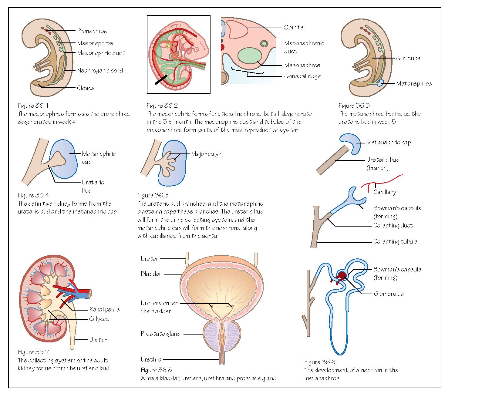
cord and the gonadal ridge (see Figure 38.2).
Three
structures involved in kidney development grow from intermediate mesoderm in an
anterior to posterior sequence, termed the pronephros, mesonephros and metanephros.
The pronephros
appears in the third week in the neck region of the embryo and disappears a
week later. In humans this is a primitive, non‐functional kidney that c ial
nephrons joined to an unbranched nephric duct.
Appearing
in the fouctional kidney unit, the mesonephros, forms as the pronephros begins
to regress (Figures 36.1 and 36.2). The mesonephric ducts (Wolffian
ducts) are epithelialined tubes that form in the intermediate mesoderm and
extend caudally to the cloaca. They stimulate formation of the mesonephros
itself as mesonephric tubules (different to the ducts) from the
mesenchyme. The tissue of the mesonephros appears initially as a segmented
structure along the mesonephric duct.
Renal
corpuscles develop from mesonephric tubules (Bowman’s capsule) and
capillaries from the dorsal aortae (glomerulus). At the lateral end the
tubules join the mesonephric duct. The duct discharges into the cloaca where
the bladder will form. The mesonephros starts to produce urine at about 6 weeks
but degenerates almost completely between weeks 7 and 10.
The
mesonephric ducts contribute to the ducts of the male reproductive system, but
regress in the female foetus (see Chapter 38).
The
third renal structure that develops will finally become the adult kidney. It
starts to appear at the beginning of the fifth week as a bud from the caudal
end of the mesonephric duct, called the ureteric bud (Figures 36.3 and
36.4).
The
bud branches and develops into the collecting parts of the adult kidney: the
ureter, renal pelvis, calyses and collecting tubules. The bud grows into
surrounding intermediate mesoderm and induces the cells in that region (the
metanephric blastema) to form a metanephric cap upon the ureteric bud.
As
the ureteric bud forms collecting tubules, cells of the metanephric cap form nephrons
that link to the collecting tubules. Reciprocal interactions between the
buds and the caps initiate and maintain this development (Figure 36.5).
Capillaries
grow into the Bowman’s capsule from the dorsal aortae and convolute to form the
glomeruli (Figure 36.6). These functional renal units produce urine from week
12 onwards.
The
formation of nephrons continues until birth when there are approximately 1
million nephrons in each kidney. Infant kidneys are lobulated because of the
branching of the calyces (Figure 36.7), but further growth and elongation of
the nephrons after birth pushes out the kidney and the lobulation disappears.
Blood supply
The
location of the metanephros changes during development from the level of the
pelvis, through growth of the embryo and migration of the kidneys, to the
lumbar region. They also rotate medially in ascent. As they ascend, a series of
blood vessels from either the common iliac arteries or aorta generate and degenerate
to continually supply the kidneys. Usually, the most cranial remain and become
the renal arteries.
In
week 4 the cloaca is split into the ventral urogenital sinus and
the dorsal anal canal by the urorectal septum (see Figure 33.5).
The
urogenital sinus can be split into a further three parts. The top part is the
biggest and becomes the bladder, the middle part forms the urethra in the
female pelvis and the prostatic and membranous urethra in the male (Figure
36.8), and the lowest part forms the penile urethra in the male and the
vestibule in the female. The allantois also contributes to the upper parts of
the bladder.
The
mesonephric ducts become incorporated into the posterior wall of the bladder.
The openings of the mesonephric ducts and ureters enter the bladder separately.
Remember that the ureters form from the metanephric ducts. The ureters move
anteriorly whereas the mesonephric ducts move posteriorly and become the
ejaculatory ducts in the male pelvis.
The
specialised transitional epithelium of the bladder develops from the
endoderm of the urogenital sinus.
The
ventral surface of the cloaca (which becomes the urogenital sinus) is
continuous with the allantois, which degenerates after birth to form the urachus
and eventually the median umbilical ligament (an embryological
remnant with no clinical significance). The medial umbilical ligaments
are the remnants of the umbilical arteries, which are a little lateral to the
urachus.
Clinical relevance
Incomplete
division of the ureteric bud can lead to supernumerary kidneys and, more
commonly, supernumerary ureters.
Kidney cysts form when the
developing nephrons fail to connect to a collecting tubule in development, or
the collecting ducts fail to develop. There are dominant and recessive forms of
polycystic kidneys. The recessive form is more progressive and often
results in renal failure in childhood.
Balance
of fluid in the amnion is vital in the development of the embryo. If urine is
not being produced there is a reduction in the amniotic fluid and oligohydramnios
develops. This can be a symptom of bilateral renal agenesis, in
which both kidneys fail to form. This is lethal. Unilateral renal agenesis
generally causes no symptoms.
Accessory renal arteries are quite
common, especially on the left and often are only seen during a surgical
procedure as they are asymptomatic. They enter the kidney at the superior and
inferior poles. Abnormal rotation or location of the kidneys may be found in a
patient, and they may fail to ascend into the abdomen. The inferior poles of
the left and right kidneys can fuse, forming a horseshoe kidney. In this case
the kidney cannot ascend as it gets snagged on the inferior mesenteric artery.
Bladder
defects may occur, such as exstrophy in which part of the ventral
bladder wall is present outside of the abdominal wall. A urachal cyst,
fistula or s the degeneration of the allantois is not completed.


