BLADDER
DUPLICATION AND SEPTATION
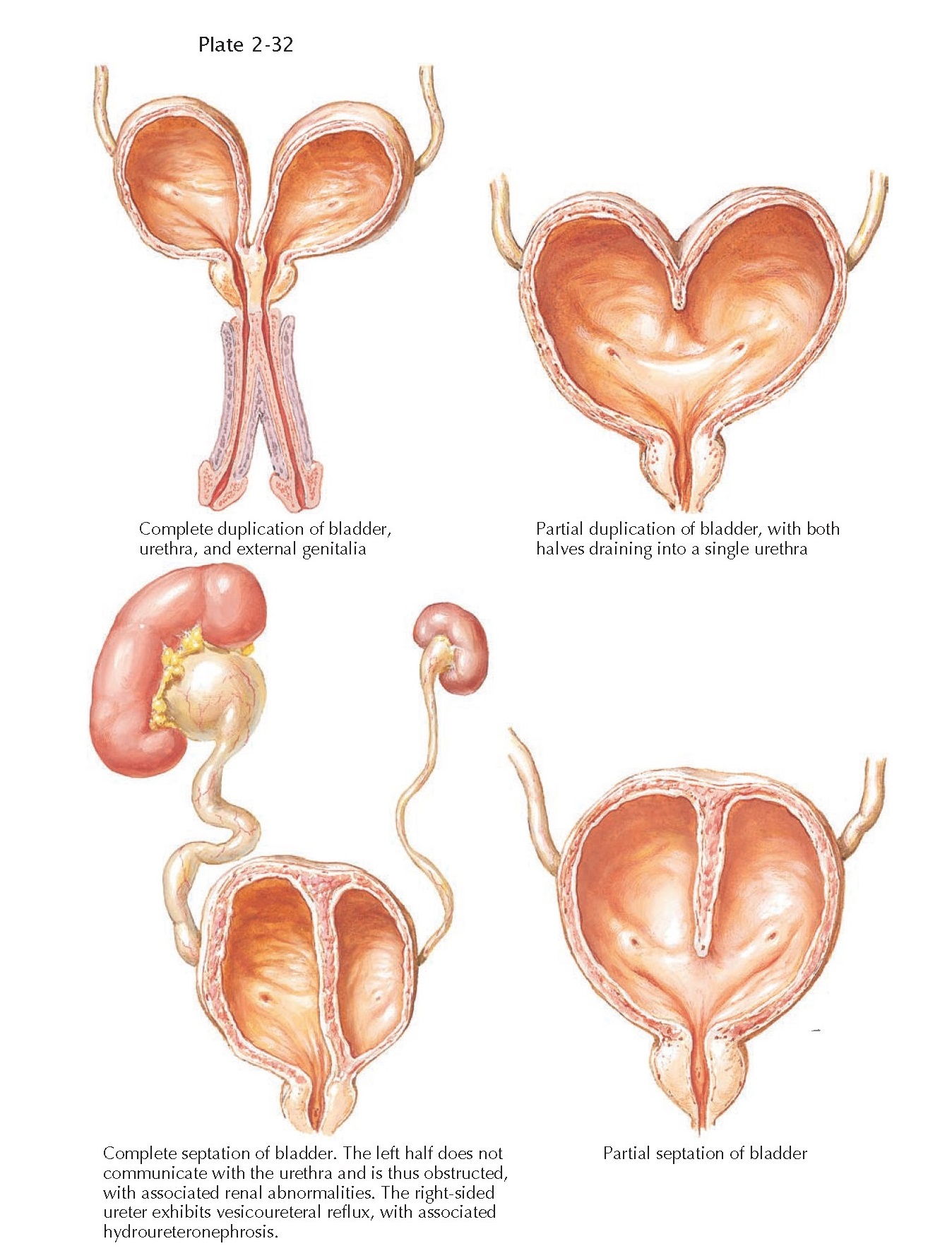
Bladder duplication and septation are very rare
congenital abnormalities, with only a small number of cases reported in the scientific
literature. In either duplication or septation, division of the bladder may be
complete or incomplete, and it may occur in the coronal or sagittal plane.
In duplication, each half of
the divided bladder receives its own ureter and possesses its own full thickness
wall. In incomplete duplication, the two halves typically unite above the level
of the bladder neck and then drain together into a single urethra. In complete
duplication, the two halves remain separate to the level of the bladder neck and
can even drain into two independent urethras, each with its own external
meatus. In some cases, however, one of the bladder halves lacks a urethral
component, resulting in outlet obstruction and ipsilateral renal abnormalities.
In septation, a fibromuscular
wall divides the bladder into separate compartments. In contrast to
duplication, septation produces two compartments that share a common wall. Like
duplication, septation can be incomplete or complete, depending on how far the
wall extends toward the bladder neck. Septation, however, is not associated
with duplication of the urethra, and thus both compartments must be in open
communication with the urethra. In some cases, however, fusion of the septum
with the bladder neck causes one compartment to lose access to the urethra,
resulting in obstruction.
Bladder duplication and
septation are frequently associated with other anomalies, especially in the
genitourinary system. For example, vesicoureteral reflux may be seen on one or
both sides, resulting in hydronephrosis if severe. Likewise, one or both of the
bladder components may lack a normal continence mechanism. If there is complete
duplication of the bladder, concurrent duplication of the external genitalia
may be seen as well. Less often, duplication may also occur in the lower
gastrointestinal tract or spine.
Pathogenesis
The embryologic basis for
these various anomalies is unknown. It is possible that complete duplication of
the bladder and adjacent organ systems reflects partial twinning of the
embryonic tail early in gestation. In contrast, isolated defects of the bladder
may reflect abnormalities during cloacal septation (see Plate 2-4).
Presentation And Diagnosis
The timing of diagnosis
depends on the nature and extent of the malformation. If there is external
evidence of duplication such as in the genitals or spine the patient is likely
to undergo comprehensive evaluation early in life, during which the bladder
abnormality will be discovered. In contrast, if there is no external evidence
of duplication, an evaluation may not be performed unless recurrent febrile
urinary tract infections occur (secondary to urine stasis and or vesicoureteral
reflux) or persistent incontinence is noted. In such cases, the bladder anomaly
should be noted during imaging with ultrasound, CT, or voiding
cystourethrography. Once the diagnosis has been established, further evaluation
should include a renal scan to assess kidney function, as well as
video-urodynamic studies to examine voiding from each bladder compartment and
to determine if vesicoureteral reflux is present.
Treatment
The need for surgical
intervention depends on the nature and extent of the malformation. If an
obstruction is present, it should be excised as soon as possible so as to
reduce the risk of further infection and preserve renal function. If incontinence,
vesicoureteral reflux, external duplication, and or other anomalies are present,
a more complex intervention will be required, the specifics of which must be
tailored to each individual patient.


