Anterior
Compartment of the Forearm Anatomy
The anterior compartment of
the forearm (Fig. 3.4) contains a superficial and a deep group of muscles,
which include flexors of the wrist, fingers and thumb and two muscles that act
as pronators. The compartment is traversed by the median and ulnar nerves and
by the radial and ulnar arteries with their venae comitantes. A layer of deep
fascia continuous with a similar layer on the posterior aspect of the limb
encloses the compartment and provides additional attachment for the superficial
muscles. In front of the carpus, deep fascia forms the flexor retinaculum (Fig. 3.30), which lies
anterior to tendons in the carpal tunnel (p. 124). Subcutaneous tissue
overlying the compartment contains cutaneous nerves and tributaries of the cephalic
and basilic veins. Branches of the medial and lateral cutaneous nerves of the
forearm may continue distal to the wrist over the carpal region of the hand.
The superficial muscles are,
from lateral to medial, pronator teres, flexor carpi radialis, palmaris longus
and flexor carpi ulnaris (Fig.
3.31). Flexor digitorum superficialis is also included in this group
but is partly covered by the other muscles.
All the superficial muscles
attach proximally to the common flexor origin on the front of the medial
epicondyle of the humerus (Fig. 3.34). In addition, pronator teres attaches to
the medial side of the coronoid process of the ulna, and flexor carpi ulnaris
attaches to the medial border of the olecranon and the adjacent part of the
subcutaneous border of the ulna. Flexor digitorum superficialis has an
additional attachment to the ulnar collateral ligament of the elbow, the
coronoid process and the anterior oblique line of the radius.
Distally, pronator teres
attaches halfway along the lateral aspect of the shaft of the radius and forms
the medial border of the cubital fossa. The muscle pronates the forearm. Flexor
carpi radialis attaches to the bases of the second and third metacarpal bones
(Fig. 3.37). It is a flexor and abductor of the wrist joint. Palmaris longus, a
vestigial muscle that may be absent, has a long thin tendon, which attaches to
the palmar aponeurosis. The muscle is a weak flexor of the wrist joint. Flexor
carpi ulnaris attaches distally to the pisiform and, via ligaments from the
pisiform, to the hook of the hamate and the base of the fifth metacarpal (Fig.
3.47). It is the most medial of the superficial muscles and is a flexor and
adductor of the wrist.
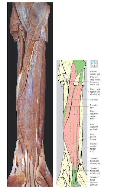
Flexor digitorum superficialis
(Fig. 3.32) is
relatively large and is the deepest of this group of muscles. Distally, it
gives rise to four tendons, one for each finger, which pass into the hand deep
to the flexor retinaculum. In the carpal tunnel the tendons have a
characteristic grouping (Fig.
3.32). Within each finger, the tendon forms two slips, which pass
around the profundus tendon and then partly reunite before attaching to the
sides of the middle phalanx (Fig. 3.37). The muscle flexes the wrist and the
metacarpophalangeal and proximal interphalangeal joints of the fingers.
The superficial muscles all
pass anterior to the elbow and therefore act as weak flexors of that joint in
addition to their roles in the movements of the wrist and hand. Collectively,
the carpal flexors and extensors stabilize the wrist joint during movements of
the fingers and thumb. Inflammation at the common flexor origin (medial
epicondylitis or Golfer’s elbow) may follow unaccustomed use of the superficial
forearm muscles and cause pain on flexion of the wrist joint.
All the superficial muscles
are innervated by the median nerve, except flexor carpi ulnaris, which is
supplied by the ulnar nerve.
Deep muscles
The deep muscles are flexor
pollicis longus, flexor digitorum profundus and pronator quadratus. Their
attachments to the radius and ulna are illustrated in Figure 3.34. Distally the
flexor pollicis longus tendon (Fig. 3.35) passes through the carpal tunnel and
attaches to the base of the distal phalanx of the thumb. The muscle flexes the
interphalangeal and metacarpophalangeal joints of the thumb. Flexor digitorum
profundus (Fig. 3.35) gives rise to four tendons, which traverse the carpal
tunnel deep to the tendons of flexor digitorum superficialis (Fig. 3.40). In
the palm, the tendons diverge, one entering each finger. Each tendon passes
between the slips of the corresponding superficialis tendon, continuing
distally to attach to the base of the terminal phalanx. The muscle is a flexor
of the fingers and of the wrist joint. Pronator quadratus, a small rectangular
muscle lying transversely between the anterior surfaces of the shafts of the
radius and ulna (Fig. 3.33),
pronates the forearm.
The deep muscles are innervated
by the anterior interosseous nerve, except for the medial part of flexor
digitorum profundus, which is supplied by the ulnar nerve.
Vessels
The brachial artery usually
divides into the radial and ulnar arteries in the cubital fossa (Fig. 3.36).
The radial artery passes distally, under brachioradialis, lying on the flexor
muscles. In the lower forearm, the vessel is accompanied by the superficial
branch of the radial nerve and, near the wrist, is sub- cutaneous and palpable
against the anterior surface of the radius. The artery winds round the lateral
aspect of the wrist, traverses the ‘anatomical snuff box’ and subsequently
enters the palm to form the deep palmar arch (Fig. 3.54). In the forearm, the
artery gives branches to muscles and contributes to anastomoses around the
elbow and wrist joints. Near the wrist, the radial artery is close to the
cephalic vein. These vessels may be joined surgically to form an arteriovenous
fistula for easy vascular access in patients undergoing renal dialysis.
The ulnar artery passes deep
to the arch formed by the radial and ulnar attachments of flexor digitorum
superficialis and continues between the superficial and deep flexor muscles. In
the distal part of the forearm, the artery is accompanied on its medial side by
the ulnar nerve. It lies beneath flexor carpi ulnaris but at the wrist emerges
to lie lateral to the tendon of this muscle, where its pulse can be palpated.
The ulnar artery crosses superficial to the flexor retinaculum and, as it
enters the hand, divides into superficial and deep palmar branches. The ulnar
artery gives branches to the muscles of the anterior compartment and to the
anastomoses around the elbow and wrist joints. Its largest branch, the common
interosseous artery (Fig. 3.36), arises near the origin of the ulnar artery and
promptly divides into posterior and anterior interosseous branches. The
posterior interosseous artery enters the posterior interosseous compartment of
the forearm (Fig. 3.77). The larger anterior interosseous artery passes distally
in the anterior compartment, lying on the interosseous membrane, accompanied by
the anterior nerve. The vessel supplies the deep flexor muscles and gives
nutrient branches to the radius and ulna. Distally, it penetrates the
interosseous membrane to assist in the anastomoses around the wrist. The
patency of the ulnar and radial arteries and of the palmar arches can be
assessed using Allen’s test. After compression of both arteries, release of one
artery should be followed within a few seconds by flushing of the whole hand.
The compression is then repeated and followed by release of the other artery.
Incomplete or slow flushing suggests poor blood flow through one of the
arteries or its branches.
Venae comitantes accompany the
arteries of the anterior compartment and drain proximally into veins around the
brachial artery.
 |
Nerves
The median nerve enters the
forearm from the cubital fossa between the two heads of pronator teres. It
crosses anterior to the ulnar artery (Fig. 3.36) and descends between the superficial
and deep flexors. At the wrist, the median nerve is remarkably superficial,
lying medial to the tendon of flexor carpi radialis and just deep to the
palmaris longus tendon. The median nerve passes through the carpal tunnel into
the hand, where it divides into terminal branches (Fig. 3.52). The nerve
supplies all the superficial muscles of the anterior compartment except flexor
carpi ulnaris. The anterior interosseous branch of the median nerve (Fig. 3.36)
supplies all the deep muscles of the compartment except the medial part of
flexor digitorum profundus. This branch lies between flexor digitorum profundus
and flexor pollicis longus and passes behind pronator quadratus to supply the
wrist (Fig. 3.33). In the forearm, the median nerve also gives a palmar
cutaneous branch, which crosses superficial to the flexor retinaculum and
supplies skin of the lateral part of the palm. Superficial lacerations near the
wrist may damage the palmar cutaneous branches but leave the median and ulnar
nerves intact. Testing digital sensation alone will miss injuries to these
nerves supplying the palm.
The ulnar nerve passes behind
the medial epicondyle where it can be palpated. Pressure here produces pain or
tingling felt in the cutaneous distribution of the nerve along the medial side
of the hand. Fractures involving the medial epicondyle may damage the ulnar
nerve. The nerve enters the forearm between the two heads of flexor carpi
ulnaris. Lying on flexor digitorum profundus and covered by flexor carpi
ulnaris, it traverses the medial side of the anterior compartment, accompanied
in the lower part of the forearm by the ulnar artery. Near the wrist, the ulnar
nerve emerges lateral to the flexor carpi ulnaris tendon and crosses
superficial to the flexor retinaculum with the ulnar artery on its lateral
side. The nerve terminates in the hand by dividing into superficial and deep
branches (p. 95). The ulnar nerve supplies the elbow joint and gives branches
to flexor carpi ulnaris and the medial part of flexor digitorum profundus. It
also provides a palmar cutaneous nerve supplying skin on the medial aspect of
the palm, and dorsal cutaneous branches that innervate the medial part of the dorsum
of the hand (Fig. 3.78).
 |
Fig. 3.36 Vessels and
nerves of the anterior compartment of the forearm. The superficial flexor
muscles and brachioradialis have been divided and most of the venae comitantes
have been removed.
|


