Spleen Anatomy
The spleen is a
lymphoid organ lying in the left hypochondrium posterior to the stomach. The
fresh spleen is purple in colour and variable in size and shape. Since it lies
entirely behind the midaxillary line and under cover of the left lower ribs,
the normal spleen cannot be palpated in the living subject, even during full
inspiration. The spleen is soft and very vascular and can be damaged by blunt
or penetrating injuries resulting in life-threatening intra-peritoneal
haemorrhage. The blood may irritate the peritoneum lining the abdominal surface
of the diaphragm, producing pain referred to the left shoulder region (p. 205).
Surface features
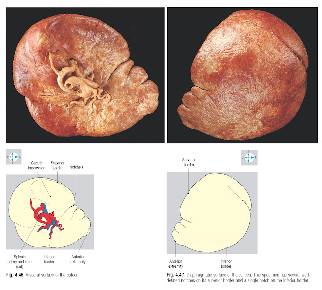
The spleen is oval in shape when viewed from its anterior
aspect (Fig. 4.46) and its long axis lies
parallel to the left tenth rib. The two extremities of the organ are connected
by superior and inferior borders. The superior border often possesses one or
more notches near its anterior end, while the inferior border is usually
smooth. The organ has two easily distinguishable surfaces. The diaphragmatic
surface faces backwards and laterally and is smoothly convex (Fig. 4.47). The visceral surface faces anteromedially and is characterized by
ridges and depressions. The centrally placed hilum is perforated by numerous
blood vessels together with lymphatics and nerves. The depressions around the hilum
accommodate adjacent organs.
Relations
The spleen is an intraperitoneal organ and most of its
capsule is covered by peritoneum of the greater sac. However, there is a small
bare area near the hilum, which gives attachment to two peritoneal folds or
ligaments. The splenorenal (lienorenal) ligament runs medially to reach the
left kidney, while the gastrosplenic ligament connects the spleen to the
greater curvature of the stomach.
Part of the
omental bursa lies between
these two ligaments and extends to the left as far as the splenic hilum
(Fig. 4.38).
Arching above the spleen and descending posterior and
lateral to it, the left dome of the diaphragm is responsible for movements of
the organ during ventilation (Fig. 4.48). The diaphragm
separates the spleen from the left lung and pleura, and from the ninth, tenth
and eleventh ribs.
On the visceral surface of the spleen, above the hilum,
is the gastric impression, which accommodates part of the posterior surface of
the stomach. Below the medial half of the hilum is the renal impression, which
abuts the superior pole of the left kidney. Near the lateral extremity of its
visceral surface, the spleen may possess a small colic impression, which lies
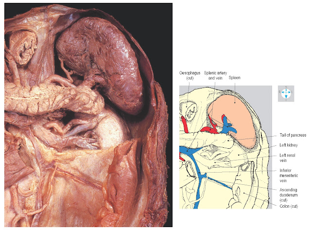
against the left colic flexure. The tail of the pancreas extends laterally into
the splenorenal ligament and its tip may reach the splenic hilum (Fig. 4.48).
 |
Fig. 4.48 Spleen and its vessels and relationship to the diaphragm,
pancreas and left kidney. The stomach and part of the colon and peritoneum have
been removed.
Blood supply
The splenic artery is a direct branch of the coeliac
trunk (p. 161). It follows a tortuous course along the upper border of the
pancreas, giving off several pancreatic branches. The artery traverses the
splenorenal ligament and divides into its terminal branches near the hilum of
the spleen. Several splenic branches enter the hilum, while the short gastric
arteries and the left gastro-omental artery enter the gastrosplenic ligament to
supply the fundus and greater curvature of the stomach, respectively.
Additional clumps of splenic tissue (splenunculi) may be present along the
course of the artery.
Veins accompany the terminal branches of the splenic
artery and unite adjacent to the hilum of the spleen to form the splenic vein.
Running to the right, this vein lies posterior to the tail of the pancreas
within the splenorenal ligament and continues retroperitoneally posterior to
the body of the gland and inferior to the splenic artery. It then crosses the
anterior aspects of the left kidney and renal vessels and receives several
small tributaries from the pancreas. Posterior to the neck of the pancreas, the
splenic vein unites with the superior mesenteric vein to form the portal vein
and, close to its termination, is usually joined from below by the inferior
mesenteric vein. If the pressure rises abnormally in the portal venous system
(portal hypertension), the spleen may become enlarged (splenomegaly).


