Nasal cavities
The
paired nasal cavities lie centrally within the facial skeleton, medial to the
orbits and the maxillary air sinuses (Fig. 7.47). They are
separated from the oral cavity by the palate, from the anterior cranial fossa
by the cribriform plates and from each other by the midline nasal septum.
Anteriorly, the cavities lead into the vestibules, which are surrounded by the
cartilaginous external nose and open onto the face at the nostrils.
Posteriorly, the nasal cavities are limited by the free edge of the nasal
septum at the choanae (posterior nasal apertures), which open into the
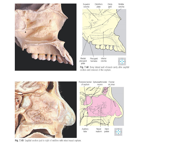
nasopharynx. Each cavity is partially subdivided by three shelf-like
projections from the lateral wall, the superior, middle and inferior conchae
(turbinates Fig. 7.48). The parts of the nasal cavity beneath each of these are
called correspondingly the superior, middle and inferior meatuses, while
above the superior concha is the sphenoeth-moidal recess. Into this recess and
the meatuses drain the paranasal air sinuses and the nasolacrimal duct.
Respiratory epithelium lines the cavity and paranasal air sinuses while the
vestibule has a stratified squamous epithelium bearing nasal vibrissae (hairs).
 |
Fig. 7.47 Coronal section showing the orbits and nasal
cavities. Posterior aspect. (Compare Figs 7.84 & 7.92.)
Bony
walls
The
medial wall is the nasal septum (Fig. 7.49), common to both cavities and formed
superiorly by the perpendicular plate of the ethmoid. This plate continues
upwards as the crista galli, which projects into the anterior cranial fossa.
The bony septum is completed posteroinferiorly by the vomer. Anteriorly, the
septum is composed of hyaline cartilage which extends into the external nose.
The
roof of each cavity comprises, from in front backwards, the nasal and frontal
bones, the cribriform plate of the ethmoid and, finally, the body of the
sphenoid bone containing the sphenoidal air sinuses. Olfactory (I) nerves from
the olfactory mucosa traverse the many small foramina in the cribriform plate
to reach the olfactory bulbs in the anterior cranial fossa (Fig. 7.50). These
nerves are vulnerable to damage in head injuries with fracture of the
cribriform plates, disrupting the sense of smell. Leakage of cerebrospinal
fluid from the nose may also result from these fractures.
The
floor of each nasal cavity is formed by the hard palate, consisting of the
palatine process of the maxilla and the horizontal process of the palatine
bone.
Numerous
bones contribute to the lateral wall (Figs 7.48,
7.50 & 7.51),
including the inferior concha and the maxilla, lacrimal, ethmoid, palatine and
sphenoid bones. The maxilla forms the
anteroinferior portion of the lateral wall and contains the maxillary air
sinus. Between the maxilla and the ethmoid, part of the lacrimal bone covers
the nasolacrimal canal, which opens into the inferior meatus. Each labyrinth
(lateral mass) of the ethmoid is attached to the lateral part of the cribriform
plate and contains numerous air cells. From the medial surface of the labyrinth
project the small superior and the larger middle conchae. The ethmoidal air
cells bulge into the middle meatus, forming the bulla, beneath which a curved
groove, the hiatus semilunaris, separates the ethmoid from the maxilla. Forming
the posterior limit of the hiatus semilunaris is the vertical plate of the
palatine bone. The most posterior component of the lateral wall is the medial
pterygoid plate of the sphenoid. Overlying the maxilla and palatine bones is a
separate bone, the inferior concha.
Sensory
nerve supply
The
somatic sensory nerve supply to the walls of the nasal cavity is derived mainly
from the maxillary (V2) division of the trigeminal nerve. The posterior lateral
nasal nerves from the pterygopalatine ganglion (p. 354) supply most of the
lateral wall, while the nasopalatine nerve supplies the septum. Lesser and
greater palatine nerves supply the posterior part of the lateral wall and the
floor. In addition, fibres from the ophthalmic (V1) division reach the nasal
cavity via the anterior ethmoidal nerve. This nerve supplies the anterosuperior parts of the
septum and the lateral wall and continues
as the external
nasal nerve to
supply the midline part of the external nose.
Most
of the blood supply to the walls of the nasal cavity is provided by branches
of the maxillary artery. These vessels arise in the pterygopalatine fossa and
are named according to the branches of the pterygopalatine ganglion they
accompany. The anteroinferior part of the nasal septum is highly vascular
(Little’s area) and commonly gives rise to nasal haemorrhage (epistaxis).
Venous
blood passes to the pterygoid plexus, the facial vein and the ophthalmic veins.
Paranasal
air sinuses
There
are four paired groups of paranasal air sinuses (Figs 7.51– 7.54) contained within the frontal, maxillary,
ethmoid and sphenoid bones. Each sinus communicates with the nasal cavity, is
lined with mucous membrane and normally contains air. The frontal air sinuses
are situated in the vertical and horizontal parts of the frontal bone, closely
related to the frontal lobes of the brain. They are variable in size and open
into the middle meatus at the infundibulum, the most anterior part of the
hiatus semilunaris. The frontal air sinus is supplied by the supraorbital
branch of the ophthalmic (V1) division of the trigeminal nerve.
The
maxillary air sinus (antrum) occupies the body of the maxilla, lying above the
oral cavity and alveolar ridge and below the orbit. Its opening at the
posterior end of the hiatus semilunaris lies high on the medial wall of the
antrum, permitting limited drainage for contents such as mucus or pus. Sensory
innervation is from the superior alveolar nerves.
The
ethmoidal air sinuses are subdivided into three groups of air cells, which
communicate with the nose through many tiny foramina. The anterior cells open
into the floor of the hiatus, while the middle cells open onto the bulla, both
groups being supplied by the anterior ethmoidal nerve. The posterior group,
innervated by the posterior ethmoidal nerve, drains into the superior meatus
under the superior concha.
The
sphenoidal air sinuses lie just below the sella turcica in the body of the
sphenoid, through the anterior wall of which they open into the sphenoethmoidal
recess. The sensory supply is from the pharyngeal branch of the pterygopalatine
ganglion. The pituitary gland can be accessed surgically through the sphenoidal
air sinus.
Infection
of the paranasal air sinuses (sinusitis) causes thickening of the mucosal
lining, which may block the openings into the nasal cavities.


