Mediastinal
Structures Anatomy
On
each side, the brachiocephalic vein is formed in the root of the neck by the
union of the internal jugular and subclavian veins. At its origin, the vein
lies behind the sternoclavicular joint and in front of the first part of the
subclavian artery.
The right brachiocephalic vein runs a
short vertical course in the superior mediastinum to unite with the left
brachiocephalic vein (Fig. 2.56) behind the medial end of the first right costal cartilage.
It receives the right vertebral and internal thoracic veins, together with the
right jugular and subclavian lymph trunks and the right lymph duct. The vessel
is accompanied by the right phrenic nerve.
The left brachiocephalic vein enters the
thorax and runs obliquely to the right, passing behind the manubrium. The
vessel lies in front of the origin from the arch of the aorta of the left
common carotid artery and the brachiocephalic trunk. At its commencement, the
vein is joined by the termination of the
thoracic duct and, along its course, receives the left vertebral, internal
thoracic and superior intercostal veins and, usually, the inferior thyroid
veins.
Superior vena cava
Formed by the union of the two
brachiocephalic veins, this large vessel descends vertically (Fig.
2.56) and terminates in
the right atrium of the heart. It lies to the right of the ascending aorta and
to the left of the right phrenic nerve and receives the azygos vein before
piercing the fibrous pericardium.
 |
Fig. 2.56 Relationships of the brachiocephalic veins to the great
arteries arising from the aortic arch.
Arch of aorta and branches
The arch of the aorta lies within the
superior mediastinum, in continuity with the ascending aorta. The vessel
curves backwards and to the left to reach the left side of the fourth thoracic
vertebral body, where it becomes the descending aorta. The arch possesses a
concavity inferiorly, left and right sides and a superior convexity.
The concavity is related to the
bifurcation of the pulmonary trunk and the left main bronchus. The ligamentum arteriosum
attaches the pulmonary trunk (or left pulmonary artery) to the concavity of the
aortic arch and is closely related to the left recurrent laryngeal nerve (Figs
2.46 & 2.57).
The left side of the aortic arch is
crossed by the left phrenic and vagus nerves (Fig. 2.57) and covered by
mediastinal pleura. The phrenic nerve lies in front of the vagus and passes
onto the fibrous pericardium in front of the lung root. The vagus nerve
inclines backwards to pass behind the lung root, having given off the left
recurrent laryngeal nerve. The left superior intercostal vein passes forwards
across the arch and usually terminates in the left brachiocephalic vein (Fig.
2.57).
The right side of the arch is related,
from in front backwards, to the superior vena cava, trachea, left recurrent
laryngeal nerve, oesophagus and thoracic duct. These structures lie between the
aorta and the right mediastinal pleura.
The convexity of the arch gives rise
to the brachiocephalic trunk, left common carotid and left subclavian arteries
(Fig. 2.58), which ascend into the root of the neck. The brachiocephalic trunk
is the first branch of the arch of the aorta and arises behind the left
brachiocephalic vein. The trunk slopes upwards and to the right across the
anterior surface of the trachea, leaving the thorax to the right of the trachea
to divide in the root of the neck into the right subclavian and right common
carotid arteries.
The left common carotid artery arises
behind the brachiocephalic trunk and ascends, in company with the left
phrenic and vagus nerves, through the superior mediastinum on the left of the trachea
into the root of the neck (Fig. 2.58).
The left subclavian artery is the most
posterior artery arising from the aortic arch and lies immediately behind the left
common carotid artery. It runs upwards and laterally, closely related to the
pleura covering the apex of the left lung, entering the root of the neck behind
the sternoclavicular joint.
The right and left phrenic nerves (C3,
C4 & C5) pass through the superior thoracic aperture behind the respective
subclavian veins. Owing to the asymmetry of the mediastinal organs, the
intrathoracic courses of the two nerves differ. The right phrenic nerve,
covered by mediastinal pleura, accompanies the right brachiocephalic vein and
the superior vena cava in front of the root of the right
lung (Fig. 2.59).
It descends vertically across the
fibrous pericardium covering the right atrium and pierces the diaphragm alongside
the infe- rior vena cava.
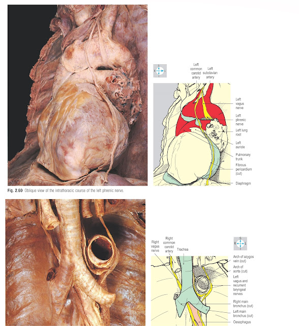
The left phrenic nerve, also covered
by mediastinal pleura, lies lateral to the left common carotid artery and
crosses the left side of the aortic arch to gain the fibrous pericardium in
front of the left lung root (Fig. 2.57). The nerve then descends across the
pericardium as far as the apex of the heart, where it pierces the diaphragm
(Fig. 2.60).
The phrenic nerves supply the muscle
of the diaphragm, excluding the crura. They give sensory fibres to the fibrous
and parietal serous pericardium and the mediastinal and diaphragmatic pleura,
and sensory branches to the peritoneum covering the inferior surface of the
diaphragm (pp 36, 205).
 |
Fig. 2.59 Oblique view showing the course of the right phrenic
nerve.
Trachea
The trachea descends through the neck, where normally it is palpable above
the jugular notch, and enters the thorax in the midline, immediately behind the
upper border of the manubrium. It runs vertically through the superior
mediastinum and, at the level of the aortic arch, divides into right and left
main bronchi (Fig. 2.61).
The right main bronchus is wider than
the left and inclines steeply downwards to enter the right lung root. The right
upper lobar bronchus often arises outside the hilum of the lung. The left main
bronchus runs obliquely to the left within the concavity of the arch of the
aorta, passing behind the left pulmonary artery to gain the left lung root.
The thoracic part of the trachea is
crossed anteriorly by the brachiocephalic trunk and the left brachiocephalic
vein (Fig. 2.59). In addition, the trachea is overlapped by the anterior margins of the
pleura and lungs and the thymus (or its remnants). The trachea is related on
the left to the arch of the aorta and left common carotid and subclavian
arteries, on the right to the superior vena cava, the termination of the azygos
vein, the right vagus nerve and the mediastinal pleura, and posteriorly to the
oesophagus and the left recurrent laryngeal nerve. (The right recurrent
laryngeal nerve does not enter the thorax but passes around the right
subclavian artery in the root of the neck; p. 331.)
The vascular supply of the trachea is
from the inferior thyroid arteries and veins. The recurrent laryngeal nerves
supply sensory and parasympathetic secretomotor fibres to the mucous membrane
and motor fibres to the smooth muscle (trachealis).
 |
Fig. 2.61 Trachea and left and right main bronchi, exposed after
removal of the anterior part of the aortic arch.
Oesophagus
The oesophagus descends through the
root of the neck and traverses the superior thoracic aperture behind the
trachea. In the superior mediastinum the oesophagus lies in front of the upper
four thoracic vertebral bodies and behind the trachea, the left main bronchus
and left recurrent laryngeal nerve. The aortic arch and the thoracic duct are
on its left while the azygos vein arches forwards on its right (Fig.
2.62).
The oesophagus continues into the
posterior mediastinum in front of the fifth thoracic vertebra accompanied by
the right and left vagus nerves. It descends behind the fibrous pericardium and
inclines to the left to cross in front of the descending aorta. On its right
side, the oesophagus is covered by mediastinal pleura. On the left, once
anterior to the descending aorta, it is related to pleura as far as the
diaphragm. Accompanied by branches of the vagus nerves (see below), the
oesophagus passes through the diaphragm at the level of the tenth thoracic
vertebra.
The oesophagus is supplied by branches
from the inferior thyroid arteries and from the descending thoracic aorta. Its
lower part receives branches from the left gastric artery that ascends through
the oesophageal opening in the diaphragm. Radicles of the left gastric vein (a
tributary of the portal vein) anastomose with veins that drain venous blood
from the oesophagus into the azygos system (see Portacaval anastomoses, p.
185). The upper part of the oesophagus is drained by the brachiocephalic veins.
Sensory and parasympathetic motor fibres to the oesophagus are provided by the
vagi and their recurrent laryngeal branches.
 |
Fig. 2.62 Intrathoracic part
of the oesophagus and accompanying vagus nerves after removal of the main
bronchi and the lower part of the trachea.
Vagus
(X) nerves
In the superior mediastinum, the
relationships of the right and left vagi differ. The right vagus nerve (Fig.
2.62) enters the thorax
behind the bifurcation of the brachiocephalic trunk and on the right of the
trachea. The nerve, covered by mediastinal pleura, inclines backwards and
passes behind the right lung root to gain the oesophagus. The left vagus nerve
descends behind the left common carotid artery to cross the left side of the
aortic arch, gives off the left recurrent laryngeal nerve and continues behind
the left lung root to reach the oesophagus.
The left recurrent laryngeal nerve (Fig.
2.62) passes around the
arch of the aorta adjacent to the ligamentum arteriosum and ascends in the
interval between the trachea and oesophagus. In the posterior mediastinum, the
right and left vagus nerves divide on the surface of the oesophagus to form a
network, the oesophageal plexus. The terminal branches of the plexus (the
anterior and posterior vagal trunks) enter the abdomen with the oesophagus (p.
197).
The descending aorta (Fig. 2.63) is
continuous with the aortic arch and initially lies to the left of the fifth
thoracic vertebral body. As it traverses the posterior mediastinum, it inclines
forwards and to the right, gaining the midline anterior to the twelfth thoracic
vertebra. On the right, the upper part of the descending aorta is related to
the thoracic vertebral bodies and the oesophagus. The lower part and all of its
left side are covered by mediastinal pleura. The thoracic duct and the azygos
vein lie to the right of the aorta, and anteriorly it is crossed by the
oesophagus sloping obliquely from the midline to the left. The descending aorta
leaves the thorax in front of the twelfth thoracic vertebra and behind the
median arcuate ligament of the diaphragm with the thoracic duct and azygos vein
(Figs 2.64 & 4.104).
Posterior intercostal arteries from
the descending aorta supply the third to the eleventh intercostal spaces on
both sides. They anastomose with the anterior intercostal arteries derived from
either the internal thoracic or the musculophrenic arteries. Other branches
from the aorta supply the right and left bronchi and the oesophagus.
Arising from the upper part of the
cisterna chyli (p. 196), the thoracic duct passes into the thorax, lying
between the azygos vein and descending aorta, and with these structures (Figs
2.62 & 2.63) ascends through the posterior mediastinum to gain the superior
mediastinum on the left of the oesophagus. The duct then curves forwards and to
the left, crossing the apex of the left lung to enter the root of the neck
where it terminates in the confluence of the left internal jugular and
subclavian veins.
Azygos venous system
This system of veins drains blood from
most of the posterior thoracic wall and from the bronchi, the pericardium and
part of the intrathoracic oesophagus. The azygos vein enters the thorax through
the aortic opening and receives posterior intercostal veins from the lower
eight spaces on the right (Fig. 2.64). Veins from the second and third spaces drain
into the right superior intercostal vein, which terminates in the azygos vein
as it arches over the right lung root to join the superior vena cava. The
venous return from the first space drains into the right brachiocephalic vein.
The azygos vein also receives the hemiazygos veins.
The hemiazygos and accessory
hemiazygos veins drain the lower eight posterior intercostal spaces on the left
side. The lowermost four spaces usually empty into the hemiazygos vein, which
crosses the midline to terminate in the azygos vein (Fig. 2.65). Veins from the next four intercostal spaces
usually join to form the accessory hemiazygos vein, which
also crosses the midline to end in the azygos. Sometimes, the hemiazygos and
accessory hemiazygos veins drain into the azygos vein by a single vessel. The
second and third spaces on the left are drained by the left superior
intercostal vein (Fig. 2.57), which crosses the aortic arch to end in the left
brachiocephalic vein. The first left intercostal space drains into the
corresponding brachiocephalic vein.
The thoracic part of the sympathetic
trunk (chain) runs along the lateral aspects of the thoracic vertebral bodies (Figs
2.65 & 2.66). In continuity with the cervical and abdominal
parts, the thoracic sympathetic trunk consists of a series of interconnected enlargements
(ganglia) occurring at intervals along
its length. Usually, each
thoracic spinal nerve is connected to its own ganglion by two branches, a
white (preganglionic) and a grey (post-ganglionic) ramus communicans. Not
infrequently, adjacent ganglia fuse together and, most often, the inferior
cervical and first thoracic ganglia fuse to form the stellate ganglion.
Branches
Fine nerve filaments running from the
sympathetic trunk contribute to the autonomic prevertebral plexuses supplying
the thoracic organs, including the heart (cardiac plexuses), lungs (pulmonary
plexuses) and the oesophagus (oesophageal plexus). The lower thoracic ganglia
give rise to a collection of autonomic fibres that form the greater (Fig.
2.66), lesser and least
splanchnic nerves, destined to supply intra abdominal structures, which are
gained by piercing the crura of the diaphragm. All thoracic spinal nerves
receive from the grey rami communicantes, sympathetic postganglionic fibres,
which are distributed to various structures of the body wall (e.g. blood
vessels, hair follicles and sweat glands) by the segmental spinal nerves.


