INTRAPULMONARY
AIRWAYS
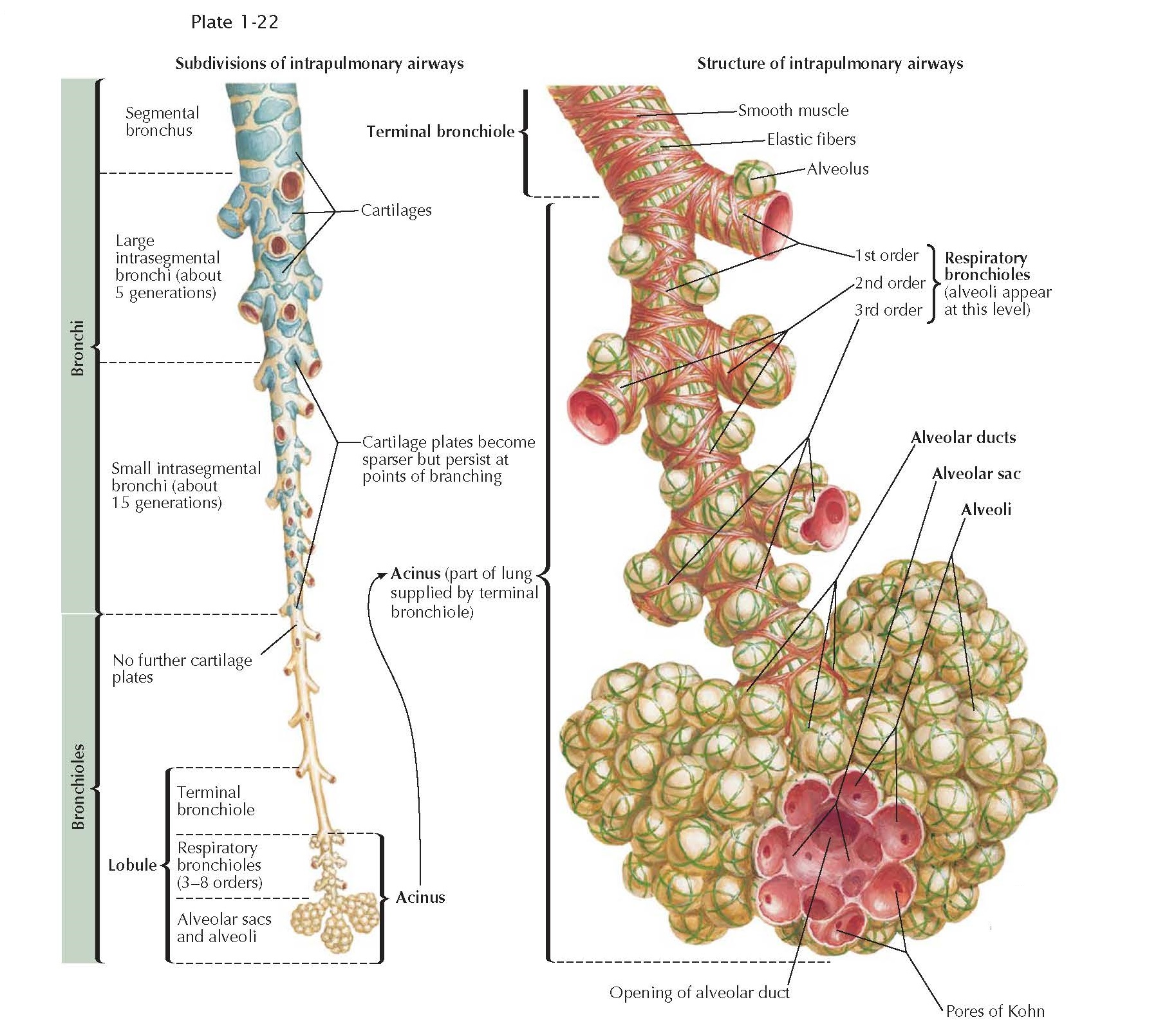
According
to the distribution of cartilage, airways are divided into bronchi and
bronchioles. Bronchi have cartilage plates as discussed earlier. Bronchioles
are distal to the bronchi beyond the last plate of cartilage and proximal
to the alveolar region. Cartilage plates become sparser toward the periphery of
the lung, and in the last generations of bronchi, plates are found only at the
points of branching. The large bronchi have enough inherent rigidity to sustain
patency even during massive lung collapse; the small bronchi collapse along
with the bronchioles and alveoli. Small and large bronchi have submucosal
mucous glands within their walls.
When any airway is pursued to its
distal limit, the terminal bronchiole is reached. Three to five terminal
bronchioles make up a lobule. The acinus, or respiratory unit, of
the lung is defined as the lung tissue supplied by a terminal bronchiole. Acini
vary in size and shape. In adults, the acinus may be up to 1 cm in diameter.
Within the acinus, three to eight generations of respiratory bronchioles may
be found. Respiratory bronchioles have the structure of bronchioles in part of
their walls but have alveoli opening directly to their lumina as well. Beyond
these lie the alveolar ducts and alveolar sacs before the alveoli proper are
reached.
None of these units is isolated from
its neighbor by complete connective tissue septa. Collateral air passage occurs
between acinus and acinus and between lobule and lobule through the pores of
Kohn in the alveolar wall and through respiratory bronchioles between adjacent
alveoli.
Connective tissue forms a sheath
around airways and blood vessels. It also forms septa that are relatively numerous
in some parts of the edges of the lingula and middle lobe and parts of the
costodiaphragmatic and costovertebral edges. These septa impede collateral
ventilation but do not prevent collateral air drift because they never com
letely isolate one unit from its neighbor in humans.


