Duodenum Anatomy
The duodenum, the
proximal portion of the small intestine, begins at the pylorus and terminates
at the duodenojejunal flexure. Deeply placed in the epigastric and umbilical
regions of the abdomen, it curves round the head of the pancreas and is shaped
like the letter ‘C’ (Fig. 4.49). Unlike the
remainder of the small intestine, the duodenum is mostly retroperitoneal and
therefore relatively immobile. The duodenal lumen receives bile and pancreatic
secretions via the bile duct and the pancreatic ducts.
 |
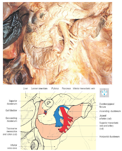
Fig. 4.49 Duodenum and some related
structures. The liver and gall bladder have been slightly displaced.
Parts and structure
The duodenum is conventionally described as consisting of
four parts (Fig. 4.49). The superior (first
part) begins slightly to the right of the midline at the level of the first
lumbar vertebra (on the transpyloric plane) and passes upwards, backwards and
to the right. In clinical practice, its initial portion is sometimes termed the
duodenal bulb or cap. The descending duodenum (second part) runs vertically to
the level of the third lumbar vertebra (Fig. 4.41). The inferior or horizontal
duodenum (third part) runs to the left across the midline, arching forwards
across the inferior vena cava and aorta. The ascending duodenum (fourth part)
slopes upwards and to the left and terminates at the level of the second lumbar
vertebra by turning sharply forwards at the duodenojejunal flexure. Close to
the pylorus the duodenal mucosa is smooth but in the second and subsequent
parts of the organ, it is raised to form numerous circular folds, the plicae
circulares (Fig. 4.50). The commonest site for duode- nal ulcers is in the
first part.
The bile duct and main pancreatic duct approach the
descending duodenum near its midpoint from the posteromedial aspect (Fig. 4.51). They usually pierce the duodenal wall in proximity and commonly open
into a single chamber, the hepatopancreatic ampulla (of Vater). The ampulla
raises a projection, the major duodenal papilla, on the internal aspect of the
duodenum. Bile and pancreatic secretions enter the duodenal lumen through the
tip of this papilla via a minute opening controlled by a ring of smooth muscle,
the ampullary sphincter (of Oddi). Immediately above the major duodenal
papilla, there is often a prominent mucosal fold forming a hood (Fig. 4.50), which may serve as a guide to the location of the papilla, particularly
during endoscopic examinations. The pancreas usually possesses a second and
smaller duct, the accessory pancreatic duct, which enters the descending
duodenum at the minor duodenal papilla, about 2 cm proximal to the major
papilla.
Relations
Most of the duodenum is retroperitoneal. However, the
initial 2 cm have peritoneal relationships similar to the stomach in that the
lesser and greater omenta attach, respectively, to the superior and inferior
borders. This short segment is relatively mobile and lies immediately inferior
to the omental foramen (Fig. 4.49). Posterior duodenal ulcers may erode the
pancreas or gastroduodenal artery (Fig. 4.52).
Anterior relations of the proximal portion of the
duodenum include the liver and gall bladder. Crossing in front of the
descending duodenum are the transverse colon and mesocolon (Fig. 4.49), below
which lie coils of jejunum and ileum. Running obliquely across the inferior
duodenum are the superior mesenteric vessels (Fig. 4.49), contained in the root
of the mesentery of the small intestine. Adjacent to the ascending duodenum are
often folds of peritoneum forming paraduodenal recesses.
Posteriorly, the superior duodenum is related to the
portal vein, the bile duct and the gastroduodenal artery (Fig. 4.52). The
descending duodenum lies in front of the hilum of the right kidney and the
right renal vessels while the inferior duodenum crosses the right ureter and
gonadal vessels, the inferior vena cava, aorta and origin of the inferior
mesenteric artery (Fig. 4.52). The ascending duodenum
ascends in front of the left psoas muscle, the left gonadal and renal vessels
and the inferior mesenteric vein.
Within the concavity of its C-shaped curve, all parts of
the duodenum are related to the pancreas (Figs 4.49 & 4.52).
Fig. 4.52 Arterial supply and some relations of the duodenum. The
superior duodenum has been displaced laterally to reveal the gastroduodenal
artery, bile duct and portal vein.
The gastroduodenal branch of the common hepatic artery
descends behind the superior duodenum and divides into right gastro-omental and
superior pancreaticoduodenal branches (Fig. 4.52). The latter vessel, which is often duplicated, runs in the interval
between the duodenum and head of the pancreas and supplies the
portion of the
duodenum proximal to
the major papilla.
The remainder of the duodenum is supplied by the inferior
pancreaticoduodenal branch of the superior mesenteric artery (Fig. 4.52), given off as the superior mesenteric artery emerges from between the
neck and uncinate process of the pancreas. The inferior pancreaticoduodenal
artery runs to the right between the duodenum and pancreas, supplying both
structures and anastomosing with the superior pancreaticoduodenal artery. The
veins draining the duodenum follow the arterial supply and terminate in the
portal venous system.


