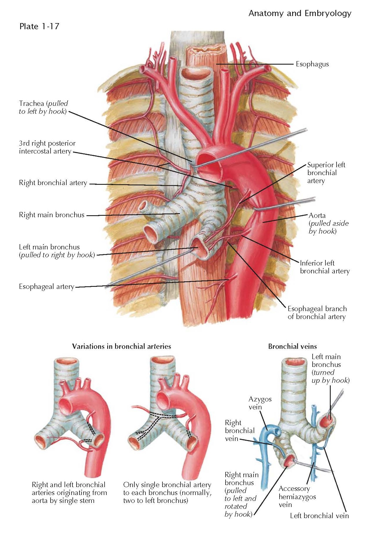
BRONCHIAL ARTERIES
The
lungs receive blood from two sets of arteries. The pulmonary arteries follow
the bronchi and ramify into capillary networks that surround the alveoli,
allowing exchange of oxygen and carbon dioxide. The bronchial arteries derive
from the aorta. They supply oxygenated blood to the tissues of the lung that
are not in close proximity to inspired air, such as the muscular walls of the
larger pulmonary vessels and airways (to the level of the respiratory
bronchioles) and the visceral pleurae. The origin of the right bronchial artery
is quite variable. It arises frequently from the third right posterior
intercostal artery (the first right aortic intercostal artery) and descends to
reach the posterior aspects of the right main bronchus. It may arise from a
common stem with the left inferior bronchial artery, which origi- nates from
the descending aorta slightly inferior to the point where the left main
bronchus crosses it. Or it may arise from the inferior aspect of the arch of
the aorta and course behind the trachea to reach the posterior wall of the
right main bronchus.
On the left side, two arteries are
typically present, one superior and one inferior. The superior artery tends to
arise from the inferior aspect of the aortic arch as it becomes the descending
aorta. The inferior artery most often arises near the beginning of the
descending aorta toward its posterior aspect. The left bronchial arteries come
to lie on the posterior surface of the left main bronchus and follow the
branching of the bronchial tree into the left lung.
Some of the more common variations of
the bronchial arteries are shown in the lower part of the illustration. The
right bronchial artery and the inferior left bronchial artery may come from a
common stem arising from the descending aorta. There may be only a single bronchial
artery on the left. Supernumerary bronchial arteries may be present, going to
either bronchus or both bronchi.
The majority of those who have studied
the blood supply of the lungs seem to agree that precapillary anastomoses are
present between the bronchial and pulmonary arteries, which can enlarge when
either of these two systems becomes obstructed (an event that more commonly
affects the pulmonary arteries). Whether these anastomoses are able to maintain
full oxygenation of an involved area of lung has not been completely
established but would seem likely given the surprisingly low rate of infarction
in otherwise normal individuals who experience pulmonary embolism.
Branches of the bronchial arteries
spread out on the surface of the lung beneath the pleura where they form a
capillary network that contributes to the pleural blood supply.


