Inguinal Canal Anatomy
The inguinal canal is
about 4 cm long and passes obliquely through the flat muscles of the abdominal
wall just above the medial half of the inguinal ligament (Fig.
4.19). In the male, the canal conveys the spermatic cord (comprising the
ductus [vas] deferens and the vessels and nerves of the testis). In the female,
the canal is narrower and contains the round ligament of the uterus.
The lateral end of the canal opens into the abdominal
cavity at the midinguinal point, defined as midway between the pubic symphysis
and the anterior superior iliac spine. In clinical practice, the midinguinal
point serves as a guide to the deep inguinal ring and the femoral artery (Fig.
4.2). There may be individual variation in the relative positions of the deep
inguinal ring, the femoral artery, and the bony landmarks, and some authors
refer to the midinguinal point or the midpoint of the inguinal ligament as
appropriate surface markings. The medial end of the canal opens into the
subcutaneous tissues at the superficial inguinal ring, an aperture in the
external oblique aponeurosis immediately superior to the pubic tubercle (Fig. 4.20). Continuous with the margins of the
superficial ring is a thin sleeve surrounding the spermatic cord, the external spermatic
fascia (Fig. 4.21).
The canal comprises a floor, a roof, and an anterior and
a posterior wall. The gutter-shaped floor is formed by the inguinal ligament (Fig. 4.22), the in-turned lower edge of the external
oblique aponeurosis. The ligament attaches laterally to the anterior superior
iliac spine and medially to the pubic tubercle and the pectineal line of the
pubis. The expanded medial end of the inguinal ligament, the lacunar ligament,
lies in the floor of the medial end of the canal, and its concave lateral edge
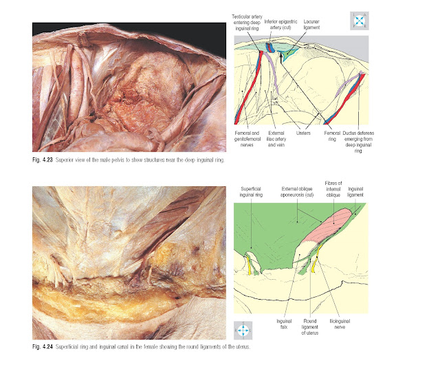
forms the medial boundary of the femoral ring (Fig. 4.23 and p. 262).
The roof is formed by the lowest fibres of internal
oblique and transversus abdominis (Fig. 4.22). These
fibres arch over the canal and pass medially and downwards to form the inguinal
falx (conjoint tendon), which attaches to the crest and pectineal line of the
pubis. The anterior wall of the canal is formed by the external oblique
aponeurosis, supplemented laterally by fibres of internal oblique. These fibres
arise from the lateral part of the inguinal ligament and cover the anterior
aspect of the deep ring (Fig. 4.21). The posterior
wall is formed by the transversalis fascia, reinforced medially by the conjoint
tendon. Deep to the transversalis fascia are the inferior epigastric vessels,
which lie just medial to the deep ring (Fig. 4.23). The inferior epigastric artery may be at risk
during operations to repair inguinal hernias.
The inguinal canal is a site of potential weakness in the
abdominal wall through which intra-abdominal structures may pass, producing an
inguinal hernia (see below). However, several features of the canal’s anatomy
minimize this weakness. The obliquity of the canal ensures that the superficial
and deep inguinal rings do not overlie one another (Fig. 4.19). Furthermore,
the strongest part of the anterior wall lies in front of the deep ring and the
strongest part of the posterior wall lies behind the superficial ring. Hence,
when pressure within the abdomen rises, the anterior and posterior walls of the
canal are firmly opposed. In addition, when the abdominal muscles contract, the
canal is compressed by the descent of fibres of internal oblique and
transversus abdominis in its roof.
Contents
In the male, the canal contains the spermatic cord (Fig.
4.21). In the female, it transmits the round ligament of the uterus (Fig. 4.24), a fibromuscular cord running from the body of
the uterus to the subcutaneous tissues of the labium majus. Lymphatics from
part of the body of the uterus accompany the round ligament and terminate in
the superficial inguinal nodes (Fig. 6.11).
In both sexes, the ilioinguinal nerve (Fig. 4.14) lies
deep to the external oblique aponeurosis close to the inguinal ligament. The
nerve runs medially in the anterior wall of the canal and emerges through the
superficial ring (Figs 4.20 & 4.24).
Inguinal hernias
The inguinal canal
is the most
common site for
an abdominal hernia. Two types of
inguinal hernia are recognized. The
direct type pushes through the inguinal falx into the medial part of the canal. By contrast, the indirect (oblique)
type traverses the deep ring and turns
medially along
the canal. Hernias
of both types may
emerge through the
superficial ring and descend
into the scrotum or labium majus.
Direct and indirect hernias are distinguished by their relationships to the inferior epigastric
vessels. A direct hernia lies
on the medial
side of
these vessels, while
the indirect type enters the inguinal canal lateral to them. The
processus vaginalis normally closes but may remain patent in infancy, leaving a
tubular channel connecting with the
peritoneal cavity. Herniation
along the patent
processus, called an infantile
inguinal hernia, is more common in the male child and may extend into the
tunica vaginalis around the testis (p. 151).


