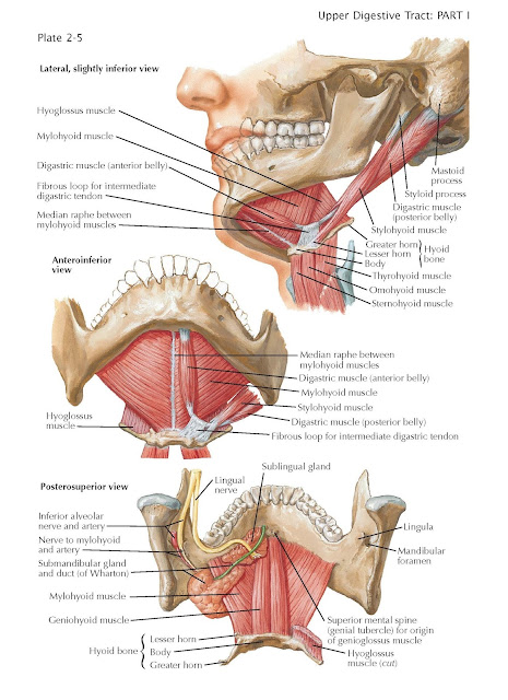
Floor of Mouth
The term floor
of the mouth is used differently by different authors, but in all cases
it is applied to the floor of the oral cavity proper and does not
include the vestibule. It is sometimes used to mean the structures that
actually serve as boundaries of the cavity inferiorly. In this sense, the
structures that form it would be the superior and lateral surfaces of the
anterior part of the tongue and the mucous membrane that is reflected from the
side of the tongue to the inner aspect of the mandible. Other authors have used
the term to mean the muscular and other structures that fill the interval
bounded by the mandible and the hyoid bone. This would mean primarily the
mylohyoid muscle, which is then thought of as the boundary between the mouth
above and the submandibular triangle of the neck below the muscle.
The right and left mylohyoid
muscles form a diaphragm that is stretched between the two mylohyoid lines
of the mandible and the body of the hyoid bone. The posterior fibers of
each muscle insert on the body of the hyoid bone, and from there forward to the
symphysis of the mandible the right and left muscles meet each other in a
midline raphe. The mylohyoid muscle is supplied by the mylohyoid nerve, which
is a branch of the inferior alveolar nerve, which itself is a branch of
the mandibular division of the trigeminal nerve.
Slightly off of the midline, the anterior
belly of the digastric muscle lies along the inferior surface of the
mylohyoid muscle. Anteriorly it attaches to the digastric fossa of the
mandible, and posteriorly it ends in the intermediate tendon, by means
of which it is continuous with the posterior belly of the digastric muscle, which
attaches to the mastoid notch of the temporal bone. The intermediate
tendon is anchored to the hyoid bone by a fascial loop. The anterior
belly is also supplied by the mylohyoid nerve and the posterior belly by a
branch from the facial nerve.
Closely related to the posterior belly
of the digastric muscle, the stylohyoid muscle extends from near the
root of the styloid process to the greater horn of the hyoid bone. It usually
attaches to the hyoid by two slips, between which the posterior belly and
intermediate tendon of the digastric muscle pass. The stylohyoid is supplied by
a branch of the facial nerve.
The right and left geniohyoid muscles,
one on each side of the midline, rest on the superior surface of the
mylohyoid muscle. They are attached anteriorly to the mental spines and
posteriorly to the body of the hyoid bone. The geniohyoid muscle is supplied by
fibers from the first cervical nerve that accompanies the hypoglossal nerve.
With the foregoing description of the
related muscles in mind, the hyoid bone can be thought of as held in a muscular
sling hung between the mandible and the stylomastoid region of the temporal
bone, thus making the floor of the mouth quite mobile. All of these muscles can
help in the elevation of the hyoid bone and the floor of the mouth. The
geniohyoid and stylohyoid muscles determine the anteroposterior position of the
hyoid bone, lengthening and shortening the floor of the mouth. The infrahyoid
(strap) muscles (omohyoid, sternohyoid, sternothyroid, and thyrohyoid)
pull the hyoid bone and floor of the mouth inferiorly.
A usage of the term floor of the
mouth which is less technical than the two previously given is to think of
the structure as the mucous membrane that is reflected from the side of the
tongue to the mandible. The attachment of the mucous membrane of this area to
the mandible, where it is continuous with the gum, is along a line drawn from
the posterior end of the mylohyoid line to a point just above the mental spine.


