Impingement Syndrome And
The Rotator Cuff
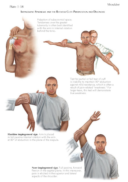
Findings associated with pathologic processes
involving the rotator cuff relate to tenderness over the rotator cuff, positive
impingement signs, and weakness of the rotator cuff demonstrated by the
internal and external rotation lag signs. Collectively, these findings are demonstrated in Plates 1-38, 1-40, and 1-43.
The Hawkins and Neer signs are
commonly called impingement signs because they are often positive when there is
inflammation, degeneration, or tears of the superior and posterior parts of the
rotator cuff. The pain associated with these physical examination signs results
from contact compression or induced strain on these parts of the rotator cuff
under the coracoacromial arch or with contact with the glenoid rim. In some
cases of shoulder pain it is not clear to the examiner if the pain is
originating from a pathologic process in the subacromial space (e.g., bursitis,
partial rotator cuff tear or full tear) when the impingement signs are
equivocal. In these cases, the examiner can perform an impingement test with
the injection of 10 mL of lidocaine or similar local anesthetic into the
subacromial space under sterile conditions. The method of injection is shown in
the later discussion on injection techniques. Several minutes after the
injection the examiner should repeat the impingement signs of the physical
examination. A positive impingement test is defined as a significant
improvement in the pain associated with these physical findings that was
present before the injection (usually 50% to 100% relief).
Chronic rotator cuff symptoms may be
progressive and symptomatic. The acromion along with the coracoid and
acromioclavicular ligament form the coracoacromial arch. In many cases these
chronic symptoms are associated with narrowing of the subacromial space or
subcoracoid space. The subacromial space is defined as the space between the
undersurface of the acromion and the rotator cuff that contains a bursa that
may become compromised in size by bone spurs that form under the acromion often
within the coracoacromial ligament. This mechanical narrowing of the space
below the coracoacromial arch can be associated with an acquired bone spur that
can cause mechanical irritation of the underlying rotator cuff. It is not
certain if the spur forms first and then causes mechanical irritation of the
rotator cuff, resulting in partial-thickness or full-thickness rotator cuff
tears or if the tear results in weakness of the rotator cuff, resulting in the
formation of a spur. In either case, the spur can be part of the pain
associated with the impingement.
Subacromial impingement and symptoms
may also occur from failure of the ossification centers of the acromion to fuse
in early adult life, resulting in a developmental anomaly called an os
acromiale. These lesions are associated with a much higher likelihood of having
a rotator cuff tear. In cases with this lesion, the tears are often larger and
occur in a younger patient population than those tears that typically occur as
a result of tendon degeneration. The most common of these types of os acromiale
is associated with lack of fusion between the anterior half and posterior half
of the acromion, result- ing in two separate bones called a meso os acromiale.
These lesions should not be confused with an acute fracture or a nonunion of an
acute fracture. The lesions can be asymptomatic. In some cases, a radiolucent
line is seen on imaging but the bone is not mobile and is associated with a
stable fibrous tissue interface. In about 60% of cases the findings are
bilateral. Because a mobile segment of bone is often tilted downward, the
anterior half of the acromion can cause mechanical irritation of the cuff and
is associated with a large tear; this more often occurs in younger patients
than the age group with degenerative or attritional tears. The os acromiale
lesions are often best seen on the axillary radiograph, with axial CT, or with
MRI. The arthroscopic removal of a lesion is generally reserved for the less
active individual. Open reduction and internal fixation with screw fixation
and, on occasion, tension band wiring is often done to treat this problem in
someone who performs heavier physical activities or heavier labor or
participates in certain sporting activities.


