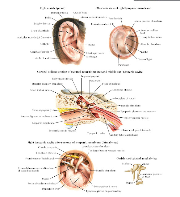
External Ear and Tympanic Cavity Anatomy
Crura of antihelix, Auricular tubercle (of Darwin), Antihelix, Concha of auricle, Lobule of
auricle, Right auricle (pinna), Triangular fossa Crux of helix, External acoustic meatus, Tragus, Antitragus, Lateral process of malleus, Posterior mallear fold, Pars flaccida, Anterior
mallear fold, Pars tensa, Handle of malleus, Umbo, Cone of light, Otoscopic view of right tympanic membrane, Tegmen tympani, Dura mater, Head of malleus, Tympanic cavity, Auditory tube
(eustachian), External acoustic
meatus, Coronal oblique
section of external acoustic meatus and middle ear (tympanic cavity),
Right tympanic cavity after removal of tympanic
membrane (lateral view), Lesser
petrosal nerve, Tympanic plexus on
promontory, Handle of malleus, Tendon of tensor tympani muscle, Lateral process of malleus, Prominence of facial canal, Long limb of incus, Chorda tympani, Malleus Incus, Lenticular process of incus, Stapes, Ossicles articulated: medial view, Intertragic notch, Scaphoid fossa, Long limb of
incus,
Pyramidal eminence and tendon
of stapedius muscle, Stapes, Tympanic nerve, Fossa of cochlear window, Long limb of incus, Footplate of
stapes, Handle of malleus, Tympanic plexus on promontory, Tensor tympani muscle, Tensor veli palatini muscle, Epitympanic recess, Superior
ligament of malleus, Short limb of incus, Chorda tympani (cut), Anterior ligment of malleus (cut), Tympanic membrane.


