BONES AND JOINTS OF
SHOULDER
The function of the upper extremity is highly dependent
on correlated motion in the four articulations of the shoulder. These include
the glenohumeral joint, the acromioclavicular joint, the sternoclavicular
joint, and the scapulothoracic articulation. The glenohumeral joint has minimal
bony constraints, thus allowing for an impressive degree of motion.
SCAPULA
Ossification centers of the scapula begin to form during the eighth week
of intrauterine life, but complete fusion does not occur until the end of the
second decade. The acromial apophysis develops from four separate centers of
ossification: the basi acromion, meta-acromion, mesoacromion, and pre-acromion.
Failure of complete fusion in a skeletally mature individual, referred to as an
os acromiale, is estimated to occur in 8% of the population, with one third of
cases being bilateral. The proximal humeral epiphysis is composed of three
primary ossification centers (the humeral head, the greater tuberosity, and the
lesser tuberosity) that coalesce at approximately age 6 years. Eighty percent
of longitudinal growth of the humerus is achieved through the proximal physis.
Physeal closure occurs at the end of the second decade.
The top of the humerus has a large, nearly spherical articular surface
surrounded at its articular margin (anatomic neck of the humerus) by two
tuberosities. The humeral head articulates with the glenoid surface, which is
only a little more than one third of its size. The great freedom of movement of
the glenohumeral joint is inevitably accompanied by a considerable loss of
stability.
The insertion of the supraspinatus portion of the rotator cuff is
superiorly on the greater tuberosity, and the infraspinatus and teres minor
insert on the posteriormost part of the greater tuberosity. All of the four
rotator cuff muscles take origin from the body of the scapula. The scapula is a
thin sheet of bone that pro- vides the site of attachment for several important
muscles of the shoulder girdle. The lateral end of the clavicle articulates with
the medial aspect of the acromion to form the acromioclavicular joint.
The large deltoid muscle has its broad origin from the spine of the
scapula posteriorly around the lateral acromion and then from the lateral third
of the clavicle. Likewise, the trapezius muscle takes its insertion over a very
similar area superior and medial to the deltoid origin. The trapezius has its
primary function in scapula retraction and elevation of scapula. The deltoid
origin on the humerus at the deltoid tuberosity is approximately one third the
distance from the shoulder to the elbow.
The levator scapulae and rhomboid major and minor insert on the medial border
of the scapula and function to retract the scapula toward the spine.
Between the anterior portion of the scapula and the chest wall (not
shown) is the scapulothoracic articulation. This articulation is another
important component of proper shoulder function. In addition to its contribution
to overall shoulder motion, rotation of the scapula brings the glenoid
underneath the humeral head so it can bear a portion of the weight of the upper extremity, thus
decreasing the necessary force generated by the muscles of the shoulder girdle.
Bony and soft tissue pathologic processes can result in bursitis and possibly
crepitus at this articulation, leading to a “snapping scapula.”
The body of the scapula has a large concavity on its costal surface, the
subscapular fossa, for the subscapularis muscle. The dorsum is convex and is
separated by the prominent spinous process into a supraspinatous fossa above,
for the supraspinatus muscle, and an infra spinatous fossa below, for the
infraspinatus muscle. The suprascapular notch is immediately medial to the coracoid
process at the superior aspect of the scapular body. The spinous process is a
large triangular projection of the dorsum of the bone, extending from the
medial border to just short of the glenoid process. It increases its elevation
and weight as it progresses laterally and ends in a concave border, the origin
of which is the neck of the scapula. The spinous process continues freely to
arch above the head of the humerus as the acromion, which overhangs the
shoulder joint. Its lateral surface provides origin for the posterior and
middle thirds of the deltoid muscle.
The coracoid process projects anteriorly and laterally from the neck of
the scapula. It gives attachment to the pectoralis minor, the short head of the
biceps brachii, the coracobrachialis, the coracoacromial ligament, and the
coracoclavicular ligaments. The lateral angle of the scapula broadens to form
the glenoid, which has minimal bony concavity. It is pear shaped, with a wider
inferior aspect. The fibrocartilaginous glenoid labrum attaches
circumferentially to the margin of the glenoid, and the long head of the biceps
brachii attaches directly to the supraglenoid tubercle.
HUMERUS
The humerus is a long bone composed of a shaft and two articular
extremities. Proximally, the head is roughly one third of a sphere, although
the anteroposterior dimension is slightly less than the superoinferior
distance. The anatomic neck is the slight indentation at the margin of the
articular surface where the capsule attaches.
The surgical neck is the narrowed area just distal to the tubercles, where
fractures frequently occur. The greater tubercle serves as the attachments for
the supraspinatus, infraspinatus, and teres minor tendons. The lesser tubercle
is the insertion of the subscapularis tendon. Each of the tubercles is
prolonged downward by bony crests, with the crest of the greater tubercle
receiving the tendon of the pectoralis major muscle and the crest of the lesser
tubercle receiving the tendon of the
teres major muscle. The intertubercular groove, lodging the long tendon of the
biceps brachii muscle, also receives the tendon of the latissimus dorsi muscle
into its floor. The shaft of the humerus is somewhat rounded above and
prismatic in its lower portion. The deltoid tuberosity is prominent laterally
over the midportion of the shaft, with a groove for the radial nerve that
indents the bone posteriorly, spiraling lateralward as it descends.
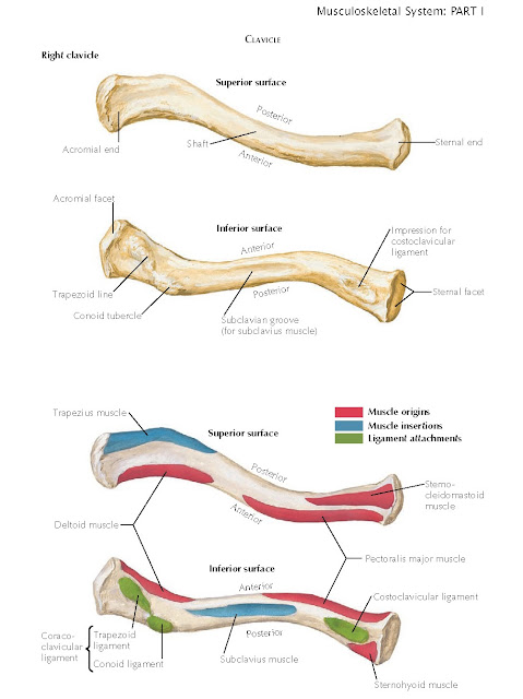
The clavicle is the first bone to ossify in the developing embryo;
however, complete ossification does not occur until the third decade of life.
When viewed from above, the clavicle has a gentle S shape with a larger medial
curve that is convex anteriorly and a smaller lateral curve that is convex
posteriorly. The medial two thirds of the bone is roughly triangular in
section, whereas the lateral third is flattened. Several bony prominences are
present on the inferior surface of the clavicle. The undersurface of the lateral
third of the bone demonstrates the conoid tubercle and trapezoid line, which
correspond to the attachment of the two parts of the coracoclavicular ligament.
Centrally, the subclavius groove receives the subclavius muscle. Medially,
there is an impression where the costoclavicular ligament attaches. The sternal
extremity of the bone is triangular and exhibits a saddle-shaped articular surface,
which is received into the clavicular fossa of the manubrium of the sternum.
The acromial extremity has an oval articular facet, directed lateralward and
slightly downward, for the acromion.
In addition to functioning as a strut that keeps the shoulder in a more
lateral position, it also serves as a point of attachment for several muscles.
Medially, the clavicular head of the pectoralis major originates anteriorly
while the sternohyoid muscle originates posteriorly. The subclavius muscle
originates from the inferior surface of the middle third of the clavicle.
Laterally, the anterior third of the deltoid originates anteriorly, a portion
of the sternocleidomastoid originates superiorly, and a portion of the
trapezius inserts posteriorly.
Resection of portions of the clavicle is typically well tolerated as long
as the integrity of the muscular attachments is not compromised. The
sternoclavicular joint represents the only true articulation between the trunk
and the upper limb. Rotation of the clavicle at this joint allows the arm to be
placed in an over-the-head position. An articular disc is interposed between
the joint surfaces, which greatly increases the capacity for movement. Joint
stability is conveyed through static stabilizers.
Stability of the shoulder is highly dependent on numerous static
stabilizers. The superior, middle, and inferior glenohumeral ligaments are
thickenings in the anterior wall of the articular capsule. Really visible only
on the inner aspect of the capsule, they radiate from the anterior glenoid
margin adjacent to and extending downward from the supraglenoid tubercle of the
scapula. These ligaments are best seen on arthroscopic photographs.
Superior Glenohumeral Ligament
The superior glenohumeral ligament (SGL) is slender, arises immediately
anterior to the attachment of the tendon of the long head of the biceps brachii
muscle, and parallels that tendon to end near the upper end of the lesser
tubercle of the humerus. The anterior biceps sling is formed by the confluence
of the SGL and the coracohumeral ligament, which stabilizes the long head of
the biceps brachii tendon as it enters the bicipital groove.
Middle Glenohumeral Ligament
The middle glenohumeral ligament (MGL) arises next to the SGL and reaches
the humerus at the front of the lesser tubercle and just inferior to the
insertion of the subscapularis muscle. It has an oblique course immediately
inferior to the opening of the subscapular bursa. When present, the middle
glenoid humeral ligament inserts on the glenoid rim posterior to the labrum.
The MGL may be cordlike, thin, or even absent. A thin middle glenohumeral ligament is seen in the arthroscopic
pictures of the shoulder allowing intra-articular visualization of most of the
articular side of the subscapularis tendon.
Inferior Glenohumeral Ligament
The inferior glenohumeral ligament arises from the scapula directly below
the notch (comma of the glenoid) in the anterior border of the glenoidal
process of the scapula and descends to the
underside of the neck of the humerus at the inferior fold
of the inferior capsular pouch. The latter two ligaments may be poorly
separated. The inferior glenohumeral ligament inserts into the anteroinferior
and posteroinferior labrum.
Coracohumeral Ligament
The coracohumeral ligament, partly continuous with the articular capsule,
is a broad band arising from the lateral border of the coracoid process.
Flattening, it blends with the upper and posterior part of the capsule and ends
in the anatomic neck of the humerus adjacent to the greater tubercle.
There are two openings in the capsule. The opening at the upper end of
the intertubercular groove allows for the passage of the tendon of the long
head of the biceps brachii muscle. The other opening is an anterior
communication of the joint cavity with the subcoracoid bursa. The synovial
membrane extends from the margin of the glenoid cavity and lines the capsule to
the limits of the articular cartilage of the humerus. It also forms the
intertubercular synovial sheath on the tendon of the biceps brachii muscle.
Coracoclavicular Ligaments
The coracoclavicular ligaments arise from the superior aspect of the base
of the coracoid. The conoid portion is more posterior and medial, whereas the
trapezoid portion is more anterior and lateral. In conjunction with the
acromioclavicular joint capsule they prevent superior displacement of the
clavicle.
Coracoacromial Ligament
The coracoacromial ligament arises from the tip of coracoid process and
attaches to the most anterior aspect of the acromion. Traction spurs may
develop at the acromial attachment, giving the acromion a more hooked shape.
This ligament plays an important role in the rotator cuff–deficient shoulder,
where it becomes the only remaining restraint to superior migration of the
humeral head.
STERNOCLAVICULAR JOINT
The sternoclavicular joint represents the only true articulation between
the trunk and the upper limb. Rotation of the clavicle at this joint allows the
arm to be placed in an over-the-head position. An articular disc is interposed
between the joint surfaces, which greatly increases the capacity for movement.
Joint stability is conveyed through
static stabilizers. The articular capsule is relatively weak but is reinforced
by the capsular ligaments. The anterior sternoclavicular ligament is a broad
anterior band of fibers attached to the upper and anterior borders of the
sternal end of the clavicle, and, below, it is attached to the upper anterior
surface of the manubrium of the sternum. This strong band is reinforced by the
tendinous origin of the sternocleidomastoid muscle. The posterior
sternoclavicular ligament has a
similar orientation on the back of the capsule and has similar bony
attachments. The costoclavicular ligament is a short, flat band of fibers
running between the cartilage of the first rib and the costal tuberosity on the
undersurface of the clavicle. The interclavicular ligament strengthens the
capsule above. It passes between the right and left clavicles with additional
attachment to the upper border of the sternum. The anterior supraclavicular
nerve gives the sternoclavicular joint its nerve supply. Blood supply is derived
from branches of the internal thoracic artery, the superior thoracic artery, and
the clavicular branch of the thoracoacromial artery.
GLENOHUMERAL JOINT
Given the lack of bony constraint, the glenohumeral joint is
circumferentially surrounded by static and dynamic stabilizers. Arthroscopic
examination of these structures is essential to accurately identify a
pathologic process in a symptomatic shoulder. The anatomic structures and their
relationship can be visualized by arthroscopy of the joint (see Plates 1-5 and
1-6). The long head of the biceps must be visualized along its entire
intra-articular course. The integrity of the biceps anchor should be examined,
as should the stability of the biceps sling at the superior aspect of the
bicipital groove. The attachment of the glenoid labrum should be inspected
circumferentially, although a sublabral foramen in the anterosuperior quadrant
can be a normal variant. An attached labrum is seen in the arthroscopic views
and art. The condition of the articular cartilage on the glenoid and humeral
head can be characterized according to its appearance on arthroscopic
examination. Grade 1 changes are seen as softening of the cartilage without
loss of the smooth cartilage surface. Grade 2 changes show loss of the smooth
cartilage surface and luster with a cobblestone appearance yet no loss of cartilage
thickness. Grade 3 indicates loss of cartilage thickness and fissuring of the
cartilage, giving it a velvet appearance when mild and the end of a mop
appearance when severe. Grade 4 is characterized by complete loss of cartilage
down to the subchondral bone. The
axillary pouch must be visualized because this is a common location of loose
bodies within the joint.
The insertion sites of the four rotator cuff tendons should be noted.
Superiorly the footprint is adjacent to the articular margin, but posteriorly
there is a bare area of bone between the articular cartilage and infraspinatus/teres
minor insertion. The subscapularis tendon is located anteriorly, and complete visualization of its insertion can
be challenging when there is a welldefined middle glenohumeral ligament. Medial
subluxation of the long head of the biceps brachii tendon from being centered
in the bicipital groove is a sign that the insertion of the subscapularis is
compromised or there is damage to the medial pulley and soft tissue wall of the biceps groove.


