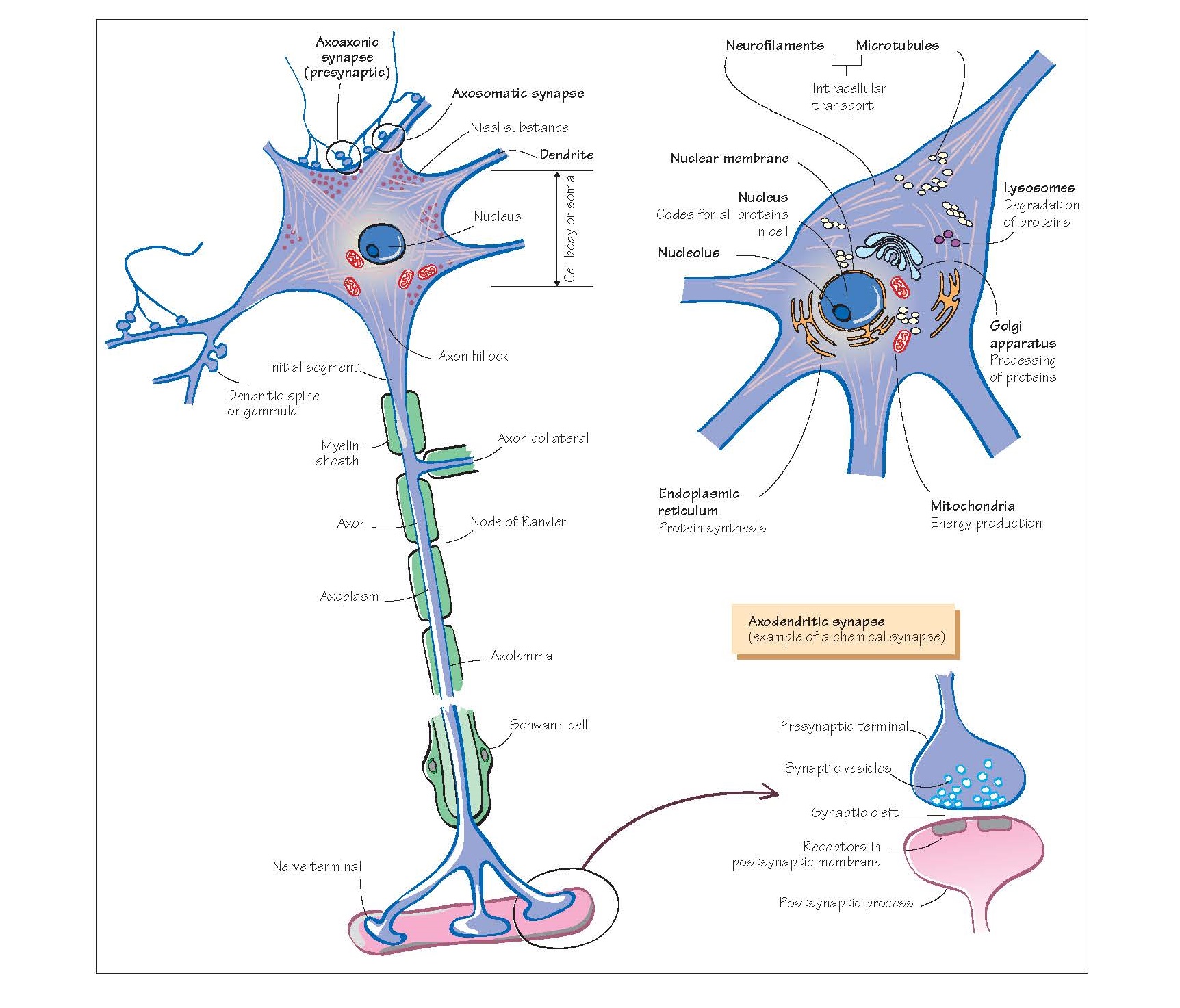
Cells Of The Nervous System I: Neurones
There are two major classes of
cells in the nervous system: the neuroglial
cells and neurones, with the latter making up only 10– 20% of the whole
population. The neurones are specialized for excitation and nerve impulse
conduction (see Chapters 14, 15 and 17), and communicate with each other by
means of the synapse (see Chapter 16) and so act as the structural and
functional unit of the nervous
system.
Neurones
The cell body (soma)
is that part of the neurone containing the nucleus and surrounding cytoplasm.
It is the focus of cellular metabolism, and houses most of the neurone’s
intracellular organelles (mitochondria, Golgi apparatus and
peroxisomes). It is typically associated with two types of neuronal processes:
the axon and dendrites. Most neurones also contain basophilic
staining, termed Nissl
substance, which is composed of granular endoplasmic reticulum and
ribosomes and is responsible for protein synthesis. This is located within the
cell body and dendritic processes but is absent from the axon hillock and
axon itself, for reasons that are not clear. In addition, throughout the cell
body and processes are neurofilaments which are important in maintaining
the architecture or cytoskeleton of the neurone. Furthermore, two other
fibrillary structures within the neurone are important in this respect:
microtubules and microfilaments, structures that are also important for
axoplasmic flow (see below) and axonal growth.
The dendrites are neuronal
cell processes that taper from the soma outwards, branch profusely and are
responsible for conveying information towards the soma from synapses on
the dendritic tree (axodendritic synapses; see also Chapter 17). Most
neurones have many dendrites (multipolar neurones) and while some inputs
synapse directly onto the dendrite, some do so via small dendritic spines or
gemmules. Thus, the primary role of dendrites is to increase the surface
area for synapse formation allowing integration of a large number of inputs
that are relayed to the cell body. In contrast, the axon, of which there
is only one per neurone, conducts information away from the soma towards the nerve
ter- minal and synapses (see Chapter 15). Although there is only one axon
per neurone, it can branch to give several processes. This branching occurs
close to the soma in the case of sensory neurones (pseudo-unipolar neurones;
see Chapter 31), but more typically occurs close to the synaptic target of the
axon. The axon originates from the soma at the axon hillock where the initial
segment of the axon emerges. This is the most excitable part of a neurone
because of its high density of sodium channels, and so is the site of
initiation of the action potential (see Chapter 15). All neurones are bounded
by a lipid bilayer (cell membrane) within which proteins are located, some
of which form ion channels (see Chapter 14); others form receptors to specific
chemicals that are released by neurones (see Chapters 18 and 19) and others act
as ion pumps moving ions across the membrane against their electrochemical
gradient, e.g. Na+–K+
exchange pump (see Chapter 15).
The axonal surface membrane is
known as the axolemma and the cytoplasm contained within it, the axoplasm.
The ion channels within the axolemma imbue the axon with its ability to conduct
action potentials while the axoplasm contains neurofilaments, microtubules and
mitochondria. These latter organelles are not only responsible for maintaining
the ionic gradients necessary for action potential production, but also allow
for the transport and recycling of proteins away from (and to a lesser extent
towards) the soma to the nerve terminal. This axoplasmic flow or axonal transport
is either slow (∼1 mm/day) or fast (∼100–400
mm/day) and is not only important
in permitting normal neuronal/synaptic activity but may also be important for
neuronal survival and development and as such may be abnormal in some
neurodegenerative disorders such
as motor neurone disease as well as disorders associated with abnormalities of certain proteins such as tau
(see Chapter 60). Many axons are surrounded by a layer of lipid, or myelin
sheath, which acts as an
electrical insulator. This myelin sheath alters the conducting properties of the
axon, and allows for rapid action potential propagation without a loss of
signal integrity (see Chapter 15). This is achieved by means of gaps, or nodes
(of Ranvier), in the myelin sheath where the axolemma contains many
ion channels (typically Na+ channels) which are directly exposed to the tissue
fluid. The nodes of Ranvier are also those sites from which axonal branches
originate, and these branches are termed axon collaterals. The myelin
sheath encompasses the axon just beyond the initial segment and finishes just
prior to its terminal arborization. The myelin sheath is formed by Schwann
cells in the PNS and by oligodendrocytes in the central nervous
system (CNS) (see Chapter 13), with many CNS axons being ensheathed by a single
oligodendrocyte while in the PNS, one Schwann cell provides myelin for one
internode.
Synapses
The synapse is the junction
where a neurone meets another cell, which in the case of the CNS is another
neurone. In the PNS the target can be muscle, glandular cells or other organs.
The typical synapse in the nervous system is a chemical one, which is
composed of a presynaptic nerve terminal (bouton or end-bulb),
and a synaptic cleft, which physically separates the nerve terminal from
the postsynaptic membrane and across which the chemical or
neurotrans-mitter from the presynaptic terminal must diffuse (see Chapter 16).
This synapse is typically between an axon of one neurone and the dendrite of
another (axodendritic synapse) although synapses are found where the
point of contact between the axon and the postsynaptic cell is either at the
level of the cell body (axosomatic synapses) or, less frequently, the
presynaptic nerve terminal (axoaxonic synapse; see Chapter 17). A few
synapses within the CNS do not possess these features but are low-resistance
junctions (gap junctions) and are termed electrical synapses. These
synapses allow for rapid conduction of action potentials without any
integration and as such tend to enable populations of cells to fire together or
in synchrony (see Chapters 16 and 61). They may also be important in the
coupling of activity across cortical areas which may be important in some of
the synchronized responses seen in the brain in sleep-wakefulness (see Chapters
43 and 44).
The specific loss of neurones is
seen in a number of neurological disorders, and those diseases in which this is
the primary event are discussed in
Chapter 60.


