The DNA molecule that stores the genetic
information in the nucleus is a long, double-stranded, helical structure. DNA
is composed of nucleotides, which consist of phosphoric acid, a five-carbon
sugar called deoxyribose, and one of four nitrogenous bases (Fig. 6.1). These
nitrogenous bases carry the genetic information and are divided into two groups:
the pyrimidine bases, thymine (T) and cytosine (C), which have one nitrogen ring, and the purine bases, adenine
(A) and guanine (G), which have two. The backbone of DNA consists of alternating
groups of sugar and phosphoric acid, with the paired bases projecting inward from
the sides of the sugar molecule.
Double Helix and Base Pairing
The native structure of DNA, as elucidated
by James Watson and Frances Crick in 1953, is that of a spiral staircase, with
the paired bases representing the steps (see Fig. 6.1). A precise complementary
pairing of purine and pyrimidine bases occurs in the double-stranded DNA molecule
in which A is paired with T and G is paired with C. Each nucleotide in a pair is
on one strand of the DNA molecule, with the bases on opposite DNA strands bound
together by hydrogen bonds that are extremely stable under normal conditions. The
double- stranded structure of DNA molecules allows them to replicate precisely by
separation of the two strands, followed by synthesis of two new complementary strands.
Similarly, the base complementary pairing allows for efficient and correct repair
of damaged DNA molecules.
Several hundred to almost 1 million
base pairs can represent a gene, the size being proportional to the protein product
it encodes. Of the two DNA strands, only one is used in transcribing the information
for the cell’s protein-building machinery. The genetic information of one strand
is meaningful and is used as a template for transcription; the complementary code
of the other strand does not make sense and is ignored. Both strands, however, are
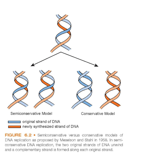
involved in DNA duplication. Before cell division, the two strands of the helix
separate and a complementary molecule is duplicated next to each original strand.
Two strands become four strands. During cell division, the newly duplicated double-stranded
molecules are separated and placed in each daughter cell by the mechanics of mitosis.
As a result, each of the daughter cells again contains the meaningful strand and
the complementary strand joined together as a double helix. In 1958, Meselson and
Stahl characterized this replication of DNA as semiconservative, as opposed
to conservative replication in which the parental strands reassociate when the two
strands are brought together (Fig. 6.2).
Packaging of DNA
The genome or total genetic content
is distributed in chromosomes. Each human somatic cell (cells other than the gametes
[sperm and ovum]) has 23 pairs of different chromosomes, one pair derived from the
individual’s mother and the other from the father. One of the chromosome pairs consists
of the sex chromosomes. Genes are arranged linearly along each chromosome. Each
chromosome contains one continuous, linear DNA helix. The DNA in the longest chromosome
is more than 7 cm in length. If the DNA of all 46 chromosomes were placed end to
end, the total DNA would span a distance of about 2 m (>6 feet).
Because of their large size, DNA molecules
are combined with several types of protein and small amounts of RNA into a coiled structure known as chromatin.
The organization of DNA into chromatin
is essential for controlling transcription and for packaging the molecule. Some
DNA-associated proteins form binding sites for repressor molecules and hormones
that regulate genetic transcription; others may block genetic transcription by preventing
access of nucleotides to the surface of the DNA molecule. A specific group of proteins
called histones is thought to control the folding of the DNA strands. Each
double-stranded DNA molecule periodically coils around histones, which keeps the
DNA organized.3 With cells that do not divide the DNA strands are in a less
compact form called chromatin. Figure 6.3 illustrates how both the chromosomes and
chromatin, which consist of chromosomal DNA, are coiled around histones.
Although solving the structural problem
of how to fit a huge amount of DNA into the nucleus, the chromatin fiber, when complexed
with histones and folded into various levels of compaction, makes the DNA inaccessible
during the processes of replication and gene expression. To accommodate these processes,
chromatin must be induced to change its structure, a process called chromatin
remodeling. Several chemical interactions are now known to affect this process.
One of these involves the acetylation of a histone amino acids group that is linked
to the opening of the chromatin fiber and gene activation. Another important chemical
modification involves the methylation of histone amino acids, which is correlated
with gene inactivation.
Genetic Code
Four bases—guanine, adenine, cytosine,
and thymine (uracil is substituted for thymine in RNA)—make up the alphabet of
the genetic code. A sequence of three of these
bases forms the fundamental triplet
code used in transmitting the genetic information needed for protein synthesis.
This triplet code is called a codon (Table 6.1). An example is the nucleotide
sequence UGG (uracil, guanine, guanine), which is the triplet RNA code for the amino
acid tryptophan. The genetic code is a universal language used by most living cells
(i.e., the code for the amino acid tryptophan is the same in a bacterium,
a plant, and a human being). Stop codons, which signal the end of a protein
molecule, are also present. Mathematically, the four bases can be arranged in 64
different combinations. Sixty-one of the triplets correspond to particular amino
acids, and three are stop signals. Only 20 amino acids are used in protein synthesis
in humans. Several triplets code for the same amino acid; there- fore, the genetic
code is said to be redundant or degenerate. For example, AUG is a
part of the initiation or start signal as well as the codon for the amino acid methionine.
Codons that specify the same amino acid are called synonyms. Synonyms usually
have the same first two bases but differ in
the third base.
DNA Repair
Rarely, accidental errors in duplication
of DNA occur. These errors are called mutations. Mutations result from the
substitution of one base pair for another, the loss or addition of one or more base
pairs, or rearrangements of base pairs. Many of these mutations occur spontaneously,
whereas others occur because of environmental agents, chemicals, and radiation.
Mutations may arise in somatic cells or in germ cells. Only those DNA changes
that occur in germ cells can be inherited.
Considering the millions of base pairs
that must be duplicated in each cell division, it is not surprising that random
changes in replication occur. Most of these defects are corrected by DNA repair
mechanisms. Several repair mechanisms exist, and each depends on specific enzymes
called endonucleases that recognize local distortions of the DNA helix, cleave
the abnormal chain, and remove the distorted region. The gap is then filled when
the correct deoxyribonucleotides, created by a DNA polymerase using the intact complementary
strand as a template, are added to the cleaved DNA. The newly synthesized end of
the segment is then joined to the remainder of the DNA strand by a DNA ligase. The
normal regulation of these gene repair mechanisms is under the control of DNA repair
genes. Loss of these gene functions renders the DNA susceptible to accumulation
of mutations. When these affect protooncogenes or tumor suppressor genes,
cancer may result.
Genetic Variability
As the Human Genome Project was progressing,
it became evident that the human genome sequence is almost exactly (99.9%) the same
in all people. It is the small variation (0.01%) in gene sequence (termed a haplotype)
that is thought to account for the individual differences in physical traits, behaviors,
and disease susceptibility. These variations are sometimes referred to as polymorphisms
(from the existence of more than one morphologic or body form in a population).
An international effort has been organized to develop a map (HapMap) of these variations
with the intent of providing a link between genetic variations and common complex
diseases such as cancer, heart disease, diabetes, and some forms of mental disease.
Mitochondrial DNA
In addition to nuclear DNA, part of
the DNA of a cell resides in the mitochondria. Mitochondrial DNA is inherited from
the mother by her offspring (i.e., matrilineal inheritance). It is a double-stranded
closed circle, containing 37 genes, 24
of which are needed for mitochondrial DNA translation
and 13 of which encode enzymes needed for oxidative metabolism. Replication of mitochondrial DNA depends
on enzymes encoded by nuclear DNA. Thus, the protein-synthesizing apparatus and
molecular components for oxidative metabolism are jointly derived from nuclear and
mitochondrial genes. Genetic disorders of mitochondrial DNA, although rare, commonly
affect tissues such as those of the neuromuscular system that have a high requirement
for oxidative metabolism.


