Cytogenetics is the study of the structure
and numeric characteristics of the cell’s chromosomes. Chromosome studies can be
done on any tissue or cell that grows and divides in culture. Lymphocytes from venous
blood are frequently used for this purpose. After the cells have been cultured,
a drug called colchicine is used to arrest mitosis in metaphase. A
chromosome spread is prepared by fixing and spreading the chromosomes on a slide.
Subsequently, appropriate staining techniques
show the chromosomal banding patterns so they can be identified. The chromosomes
are photographed, and the photomicrographs of each of the chromosomes are cut out
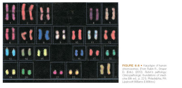
and arranged in pairs according to a standard classification system (see Fig. 6.6).
The completed picture is called a karyotype, and the procedure for preparing
the picture is called karyotyping. A uniform system of chromosome
classification was originally formulated
at the 1971 Paris
Chromosome Conference and was later revised
to describe the chromosomes as seen in more elongated prophase and pro-metaphase
preparations.
In the metaphase spread, each chromosome
takes the form of chromatids to form an “X” or “wishbone” pattern. Human chromosomes
are divided into three types according to the position of the centromere. If the
centromere is in the center and the arms are of approximately the same length, the
chromosome is said to be metacentric; if it is not centered and the arms
are of clearly different lengths, it is submetacentric; and if it is near
one end, it is acrocentric. The short arm of the chromosome is designated
as “p” for “petite,” and the long arm is designated as “q” for no other reason than
it is the next
letter of the alphabet. The arms of the chromosome
are indicated by the chromosome number followed by the p or q designation (e.g.,
15p). Chromosomes 13, 14, 15, 21, and 22 have small masses of chromatin called
satellites attached to their short arms by narrow stalks. At the ends of
each chromosome are special DNA sequences called telomeres. Telomeres allow
the end of the DNA molecule to be replicated completely.
The banding patterns of a chromosome
are used in describing the position of a gene on a chromosome. Each arm of a chromosome
is divided into regions, which are numbered from the centromere outward (e.g.,
1, 2). The regions are further divided into bands, which are also numbered (Fig.
6.9). These numbers are used in designating the position of a gene on a chromosome.
For example, Xp22 refers to band 2, region 2 of the short arm (p) of the X chromosome.


