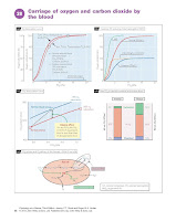
Carriage Of Oxygen
And Carbon Dioxide By The Blood.
Oxygen
The resting O2 consumption in adults
is ∼250 mL/min, rising to
>4000 mL/min during heavy exercise. The
O2 solubility in plasma is, however,
low and at a Po2 of 13 kPa blood contains
only 3 mL/L of dissolved O2 in solution. Most O2 is therefore carried bound to haemoglobin in
red blood cells. Each gram of haemoglobin can combine with 1.34 mL of O2 and so, for a haemoglobin concentration
[Hb] of 150 g/L, blood can contain a maximum of 200 mL/L of O2 (O2 capacity).
The actual amount of O2 bound to haemoglobin (O2 content) depends on the
Po2, and the percentage O2 saturation = content/ capacity × 100 (Fig. 28a).
Each haemoglobin molecule binds up to four O2 molecules; binding is cooperative,
so that the binding of each O2 molecule makes it easier for the next. This steepens
the O2 hae moglobin dissociation curve, which describes
the relationship between blood O2 content and Po2 (Fig.
28a). The curve flattens above ∼8 kPa Po2
as all binding sites become occupied. Thus, for a normal arterial Po2 (∼13 kPa) and [Hb], the blood
is ∼97% saturated and contains slightly less than 200 mL/L of O2. Because the
dissociation curve is flat in this region, any increase in Po2 (breathing O2-enriched
air) will have little effect on content. On the steep part of the curve,
however (<8 kPa Po2), small changes in Po2 will have large effects on
content.
Oxygen uptake and delivery. The high PO2 in the lungs facilitates O2 binding
to haemoglobin, whereas the low Po2 in the tissues encour-ages release. The dissociation
curve is shifted to the right (reduced affinity, facilitating O2 release) by a fall
in pH, a rise in Pco2 (Bohr shift) and an increase in temperature, which
occur in active tissues (Fig. 28a). The metabolic by-product 2,3-diphosphoglycerate
(2,3- DPG) also causes a right shift. In the lungs, Pco2 falls, the pH consequently
rises and the temperature is reduced; these all increase affinity and shift the
curve to the left, facilitating O2 uptake.
Anaemia. This is an abnormally low [Hb]; the O2 capacity
is there-fore less and the O2 content at any Po2 is reduced (Fig. 28b). Arterial
Po2 and O2 saturation remain normal. In order to deliver the same amount of O2 to
the tissues, the capillary Po2 would have to fall further than normal (Fig. 28b),
reducing the driving force for O2 diffusion into the tissues. The latter may become
inadequate for metabolism, especially during exercise, although a 50% reduction
in [Hb] does not usually cause symptoms at rest.
Carbon monoxide. Carbon monoxide (CO) binds 240 times more
strongly than O2 to haemoglobin and, by occupying O2-binding sites, reduces the
O2 capacity. However, unlike anaemia, CO also increases the affinity and shifts
the dissociation curve to the left, making O2 release to the tissues more difficult.
Thus, if 50% of haemoglobin is bound to CO, Po2 needs to fall much further than
in anaemia to release the same amount of O2, causing symptoms of severe hypoxia
(head- ache, convulsions, coma, death) (Fig. 28b).
Fetal haemoglobin. Fetal haemoglobin (HbF) binds 2,3-DPG less
strongly than does adult haemoglobin (HbA), and so the dissociation curve is shifted
to the left. This facilitates the transfer of O2 from maternal blood to the fetus, where the arterial Po2 is only ∼5 kPa (Fig. 28b).
Carbon dioxide
CO2 is formed in the tissues and transported
to the lungs where it is expired. Blood can carry much more CO2 than O2, as can be seen in the CO2 dissociation curve (Fig. 28c). This is also
more linear than the O2 dissociation
curve and does not plateau. CO2 is transported as bicarbonate, carbamino compounds
and simply dissolved in plasma (Fig. 28d).
Bicarbonate. Approximately 60% of CO2 is carried as bicarbonate. Water and CO2 combine to form carbonic acid (H2CO3) and thence
: CO + H O ⇔ H CO ⇔ HCO− + H+. The left bicarbonate (HCO3) side of the equation is normally
slow, but speeds up dramatically in the presence of carbonic anhydrase, found
in red cells. Bicarbonate is therefore formed preferentially in red cells, from
which it easily diffuses out. Red cells are, however, impermeable to H+ ions, and
Cl− enters the cell to maintain electrical
neutrality (chloride shift) (Fig.
28e). H binds avidly to deoxygenated
(reduced) haemoglobin (haemoglobin
acts as a buffer), and so there is little increase in [H+] to impede further bicarbonate
formation. Oxygenated haemoglobin does not bind H+ as well, and
so in the lungs H+ dissociates from
haemoglobin and shifts the CO2–HCO− equation to the left, assisting CO2 unloading from the blood (Fig.
28e); the reverse occurs in the tissues. This contributes to the Haldane effect,
which states that, for any Pco2, the CO2 content of oxygenated
blood is less than that of deoxygenated blood. Thus the red line A–X in Figure 28c
shows the relationship between CO2 content and PCO2 if the
blood remained 98% saturated with O2. Mixed venous O2 saturation
is however ∼75%, so as the blood becomes oxygenated in the lungs or deoxygenated in
the tissues, the relationship between CO2 content and Pco2 actually
follows the dashed line A–V.
Carbamino compounds. These compounds are formed by the reaction of
CO2 with protein amino groups: CO2 + protein-NH2 ⇔ protein-
NHCOOH. The most prevalent protein in blood is haemoglobin, which forms carbaminohaemoglobin
with CO2. This occurs more readily for deoxygenated than oxygenated
haemoglobin, contributing to the Haldane effect (Fig. 28c). Carbamino compounds
account for 30% of CO2 carriage.
Dissolved carbon dioxide. CO2 is 20 times more soluble than O2 in
plasma, and ∼10% of CO2 in blood is
carried in solution.
Hyperventilation and hypoventilation
Doubling the rate of ventilation halves
the alveolar and arterial Pco2. Ventilation is normally closely matched
to the metabolic rate as reflected by CO2 production (Chapter 29). Hyperventilation
(over- ventilation) and hypoventilation (underventilation) are defined
in terms of arterial Pco2, so that a subject is hyperventilating when
Pco2 is <5.3 kPa, and
hypoventilating when Pco2 is >5.9 kPa. Rapid breathing in exercise
is not hyperventilation,
as this is
appropriate for increased CO2 production and Pco2 does not fall. Hyperventilation
cannot normally increase the O2 content, as arterial haemoglobin is
already nearly fully saturated. The fall in Pco2 (hypocapnia) during
hyperventilation causes light-headedness, visual disturbances due to cerebral vasoconstriction
(Chapter 24) and muscle cramps (tetany). Hyperventilation can be caused by pain,
hysteria and strong emotion. Hypoventilation causes a high Pco2 (hypercapnia)
and a low Po2 (hypoxia), and may be caused by head injury or respiratory
disease.


