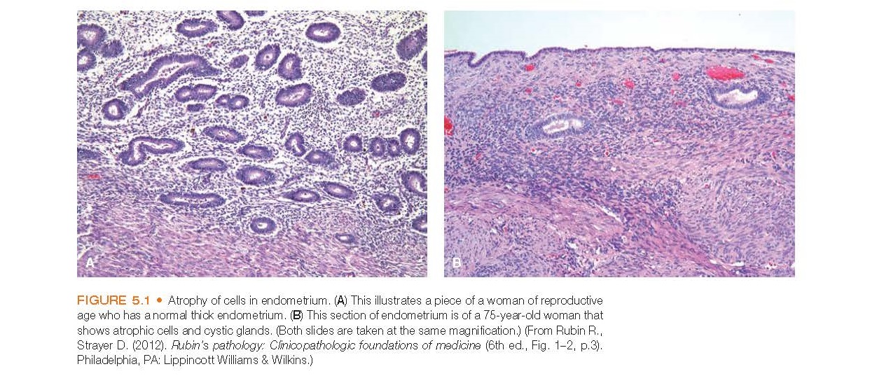
When confronted with a decrease in
work demands or adverse environmental conditions, most cells are able to revert
to a smaller size and a lower and more efficient level of functioning that is
compatible with survival. This decrease in cell size is called atrophy and
is illustrated in Figure 5.1 regarding atrophy of the endometrium. Cells that
are atrophied reduce their oxygen consumption and other cellular functions by
decreasing the number and size of their organelles and other structures. There
are fewer mitochondria, myofilaments, and endoplasmic reticulum structures.
When a sufficient number of cells are involved, the entire tissue or muscle
atrophies.
Cell size, particularly in muscle
tissue, is related to work-load. As the workload of a cell declines, oxygen
consumption and protein synthesis decrease. Furthermore, proper muscle mass is
maintained by sufficient levels of insulin and insulin-like growth factor-1
(IGF-1). When insulin and IGF-1 levels are low or catabolic signals are
present, muscle atrophy occurs by
mechanisms that include
reduced synthetic processes, increased proteolysis by the
ubiquitin–proteasome system, and apoptosis or programmed cell death. In the
ubiquitin–protea-some system, intracellular proteins destined for destruction
are covalently bonded to a small protein called ubiquitin and then
degraded by small cytoplasmic organelles called proteasomes. The general
causes of atrophy can be grouped into five categories:
•
Disuse
•
Denervation
•
Loss of
endocrine stimulation
•
Inadequate
nutrition
•
Ischemia
or decreased blood flow
Disuse atrophy occurs when there is
a reduction in skeletal muscle use. An extreme example of disuse atrophy is
seen in the muscles of extremities that have been
encased in plaster casts. Because atrophy is adaptive and reversible, muscle
size is restored after the cast is removed and muscle use is resumed.
Denervation atrophy is a form of disuse atrophy that occurs in the muscles of
paralyzed limbs. Lack of endocrine stimulation produces a form of disuse
atrophy. In women, the loss of estrogen stimulation during menopause results in
atrophic changes in the reproductive organs. With malnutrition and decreased
blood flow, cells decrease their size and energy requirements as a means of survival.


